
Overview
Aster Whitefield Hospital stands at the forefront of cutting-edge neurosurgical care, addressing a wide spectrum of conditions affecting the brain. Our team boasts of over 70 years of collective expertise and has established a leadership position in the region, delivering patient outcomes that meet global standards of excellence.
The Aster International Institute of Neurosciences exemplifies our dedication to advanced neurosurgical care. Staffed with a multidisciplinary team of highly skilled neurosurgeons, neurologists, neuro-anaesthetists and other allied specialists we leverage the latest diagnostic and treatment technologies to provide exceptional care.
Highlights of Aster Whitefield for neurosurgical care:
Expert Team: Highly experienced and qualified neurosurgeons.
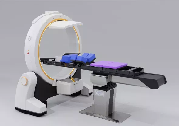
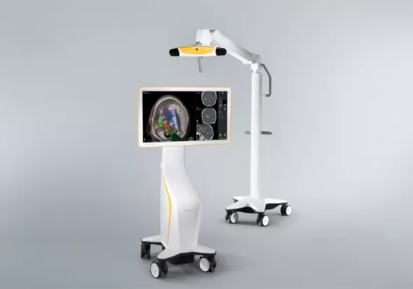
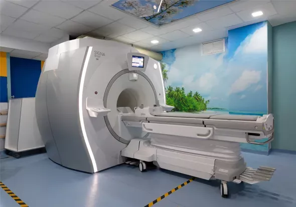
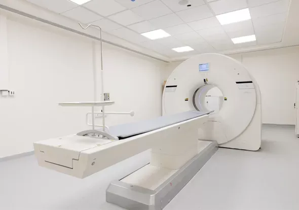
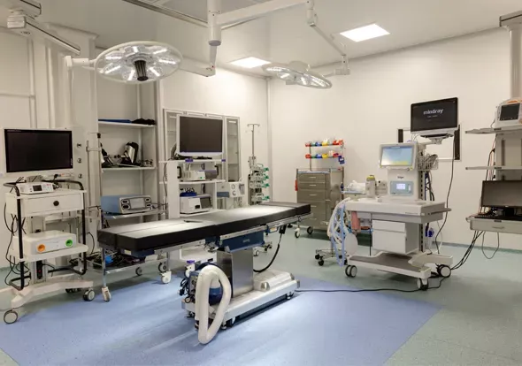
State-of-the-art technology: Latest advancements in medical technology for precise diagnosis and effective treatment.
Comprehensive Treatment Range: Wide array of treatment options for various neurosurgical conditions.
Commitment to Patient Care: Dedication to patient well-being ensures comprehensive and compassionate care.
Rehabilitation Services: Extensive rehabilitation services to support recovery and help patients regain their normal lives post-surgery.
At Aster Whitefield Hospital, we are committed to delivering holistic and integrated care. Our neurosurgical team collaborates closely with other medical specialists to ensure that every patient receives comprehensive, multidisciplinary care. For those seeking top-tier neurosurgical services in India, Aster International Institute of Neurosciences represents an excellent choice, blending advanced medical expertise with a steadfast commitment to patient-centered care.
At Aster Whitefield Hospital, our Neurosurgical Care Centre provides patient-centric brain tumor treatments. Our team of highly skilled neurosurgeons utilize advanced surgical techniques to ensure the best possible outcomes for our patients.
Our approach to brain tumor surgery incorporates several cutting-edge techniques like functional MRI, neuro-navigation, intraoperative neuromonitoring, ultrasound, real-time ultrasound and fluorescence technology to aid in tumour removal
These sophisticated techniques allow our surgeons to perform brain tumor surgeries with maximum safety and effectiveness. Our department specializes in treating all types of brain tumors, from benign meningiomas to malignant gliomas.
An acoustic schwannoma, also known as a vestibular schwannoma or acoustic neuroma, is a non-cancerous tumour that develops on the vestibular nerve, which connects the inner ear to the brain. Diagnosis begins with a thorough clinical evaluation, including a detailed medical history and physical examination to identify symptoms such as hearing loss, tinnitus, and balance issues. Audiometric tests assess hearing loss while imaging studies like MRI with gadolinium contrast are used to visualize the tumor. CT scans and electrophysiological tests, including auditory brainstem response (ABR) may be done for further assessment. Surgery is indicated for larger tumours or those causing significant symptoms.
A brain tumor is an abnormal growth or mass of cells in the brain. These tumours can be either benign or malignant. A craniotomy is a surgical procedure where a portion of the skull bone is removed to access the brain depending on the location of the tumour. Neuronavigation is used to plan the site of entry and the size of the opening depending on the preoperative images of the patient. The tumour will be identified and removed based on its location within the brain.
Intraventricular tumors are growths that develop within the brain’s ventricles, which are hollow spaces filled with cerebrospinal fluid. Surgery is a primary treatment option for intraventricular tumors, tailored to the tumor’s type, size, location, and the patient’s overall health. We offer two main surgical approaches namely Minimally Invasive Endoscopic Surgery and Craniotomy.
A pituitary tumor or adenoma is an abnormal mass or growth that occurs in the pituitary gland, a small pea-sized gland in the brain. This gland produces hormones that regulate the functions of various other glands. Although most pituitary tumors are not cancerous, they can disrupt hormone production, leading to various health issues in the body. Medications are prescribed to control the symptoms, hormonal secretion or to shrink the tumors. Surgery is done if the tumor causes symptoms such as vision problems or alters hormone levels. Craniotomy is preferred in case of large tumor masses
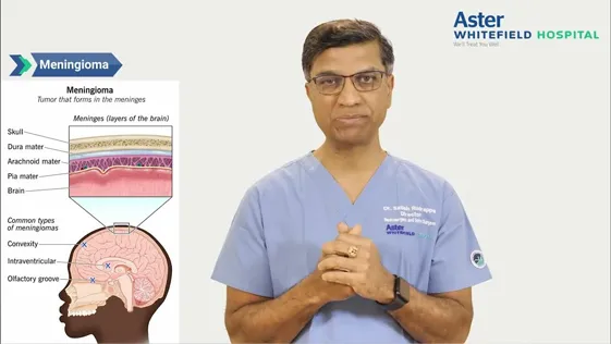
Most meningiomas are benign and do not invade other organs. They originate in the Arachnoid of the meninges and are more common in the brain. We employ advanced diagnostic techniques to accurately identify meningiomas. Utilizing MRI and CT scans we determine the presence, size and precise location of the tumours. In some cases, a biopsy is performed to confirm the diagnosis and assess the tumor type. Comprehensive neurological examinations are conducted to evaluate the impact on brain functions and advanced imaging techniques such as PET scans provide detailed tumor mapping.
Our approach to meningioma surgery includes meticulous preoperative planning with 3D imaging to map out the tumor and determine the best surgical strategy. We utilize minimally invasive techniques to reduce recovery time and minimize surgical risks.
Glioma is a type of brain tumor. Patients suspected of having gliomas undergo a comprehensive set of tests to confirm the diagnosis and determine the tumor's grade which includes clinical examination, MRI scans and sometimes a biopsy if required. Treatment plans are tailored based on the tumor's grade, location, and the patient's overall health.
Craniotomy or an open brain surgery technique is most commonly used for glioma. In cases where the tumor is in proximity to crucial brain regions, surgery may be complemented by intraoperative MRI or intraoperative brain mapping.
For chordomas, our treatment approach typically includes a combination of surgical resection and radiation therapy. Despite the challenges, our goal is to achieve optimal tumor control and improve the quality of life for our patients.
Trigeminal schwannomas are often addressed through microsurgical techniques aimed at complete tumor removal. While these procedures can be associated with high morbidity, our experienced surgeons utilize advanced techniques to minimize risks and maximize recovery.
Craniosynostosis involves the premature fusion of skull sutures, leading to abnormal skull development. It occurs when the growth seams in the skull fuse too early. Craniosynostosis is usually treated with surgery to unlock the fused bones and reshape the skull. Our neurosurgical team has the highest experience in the treatment of such complex conditions
This minimally invasive surgery implants thin electrodes near specific areas within the brain responsible for movement control. A pacemaker-like device placed under the chest sends electrical pulses to these electrodes, regulating abnormal activity and improving symptoms of Parkinson's disease, tremors, dystonia (muscle contractions), and even some chronic pain conditions. The settings of the stimulator can be adjusted by a doctor to optimize symptom control.
This minimally invasive procedure that involves placing coils or sometimes flow divertors to block blood flow and prevent rupture.
During an endovascular procedure, a flexible catheter is introduced through an artery in the groin or wrist and directed to the brain aneurysm under fluoroscopy guidance. Soft platinum wire coils are typically inserted into the aneurysm, inducing clot formation to obstruct blood flow. Alternate materials like stents or flow diverters may be employed for differently shaped or sized aneurysms.
A neurosurgeon performs a craniotomy to access the brain, followed by placing a metal clip at the base of the aneurysm, effectively sealing it off from the surrounding blood vessels and preventing rupture. This method offers a permanent solution and is particularly suitable for certain types of aneurysms or patients who are not suitable for endovascular treatment.
Preferred for many intraventricular tumors, this technique involves making a small incision in the scalp. A neurosurgeon inserts a thin tube equipped with a camera and light to visualize and remove the tumor using specialized instruments. This method is less invasive than traditional surgery, promoting quicker recovery.
It is a minimally invasive neurosurgical technique that uses microscopes and precision instruments to carefully remove or repair tissues, often in delicate areas of the brain.
This technique uses stent-like devices to divert blood flow away from the aneurysm. Over time, this allows the aneurysm to heal naturally.
Sometimes when the size or/ and the shape of an aneurysm is not suitable for either endovascular treatment or standard surgical clipping a bypass of the involved segment of the blood vessel is done to prevent the aneurysm from rupturing.
For larger or more complex tumors, traditional open brain surgery may be necessary. This involves making a larger incision in the scalp and temporarily removing a section of the skull to access and remove the tumor. Post-surgery, the skull and scalp are carefully repaired.

This advanced diagnostic and surgical infrastructure allows the Department of Neuroscience at Aster Whitefield Hospital to provide patients with exceptional neurological care. By utilizing these tools, our team can achieve a deeper understanding of each patient's condition, leading to more effective treatment plans and improved patient outcomes.


Blogs