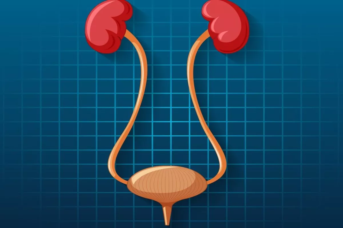
Urology is a branch of medicine that deals with the diagnosis and treatment of conditions related to the urinary tract and male reproductive system. The urology department of Aster Whitefield Hospital is one of its vital departments, which caters to patients with a wide range of urological conditions, including urinary tract infections, kidney stones, prostate cancer, bladder cancer, erectile dysfunction, infertility, and more.
The urology department is an indispensable part of Aster Whitefield Hospital, providing essential services to patients with a wide range of urological conditions. With advanced diagnostic tools, skilled healthcare professionals, and personalized treatment plans, our urology department is committed to providing patients with the best possible care and outcomes. The urology department of Aster Whitefield Hospital has a team of highly trained and experienced urologists, nurses, and support staff who work together to provide excellent patient care. The department is equipped with the latest technology and equipment, including state-of-the-art diagnostic tools such as ultrasound, CT scans, MRI machines, and much more to diagnose and treat urological conditions effectively.
The first step in the treatment of any patient by our urology department is to conduct diagnostic tests and procedures to determine the underlying cause of a patient's symptoms. These may include urine tests, blood tests, biopsies, and imaging studies. Based on the test results, the urology team develops an individualized treatment plan, which may include medication, surgery, or other interventions.
Our urology department also plays a crucial role in providing preventative care, such as routine prostate exams and testicular exams for men, and regular pelvic exams and Pap tests for women. By detecting urological conditions we ensure that the patients have a higher chance of successful treatment and recovery.
In addition to diagnosing and treating urological conditions, the urology department of Aster Whitefield also provides patient education and support. Patients are often encouraged to make lifestyle changes, such as dietary modifications and exercise, to improve their overall health and reduce their risk of urological conditions.
Services We Offer
Some of the major services offered by our urology department include;
Evaluation, diagnosis, and treatment of urological conditions: Our Urology team offers a wide range of services to evaluate and treat urological conditions such as urinary tract infections, kidney stones, prostate problems, bladder issues, sexual dysfunction, renal transplant, infertility, urinary incontinence, bladder prolapse.
Surgical procedures: Our bright, eager-minded urologists with high precision dexterity and skills perform a variety of surgical procedures, including minimally invasive and robotic surgeries. Common surgeries performed by our urologists include;
- Renal transplants
- Prostatectomies
- Bladder surgeries
- kidney stone removal
- Open Urological Surgery
- Holium Laser for calculus and prostrate surgery
- Vasectomies
- Penile Plication
- Penile implants and prosthesis
- Circumcision.
Cancer care: Our Urology Department has specialists who treat various types of urological cancers, such as prostate cancer, bladder cancer, kidney cancer, and adrenal gland cancer. Our specialists are highly trained in the latest techniques in cancer treatment, such as radiotherapy, chemotherapy, and cryosurgery. They are also proficient in minimally invasive treatments, such as robotic and laparoscopic surgery.
Pediatric urology: Pediatric urology is a specialized field of medicine that focuses on diagnosing and treating urological conditions in children. This includes situations that affect the kidneys, bladder, and other parts of the urinary system. It also includes birth defects related to the urinary system. We evaluate and treat children with urological conditions such as bedwetting, urinary tract infections, and congenital abnormalities
Incontinence treatment: Our Urology Department offers treatment options for urinary incontinence, including medications, behavioral therapy, and surgical procedures. The medications work by relaxing the bladder, increasing bladder capacity, or reducing bladder spasms. Behavioral therapy helps to retrain the bladder muscles and help with urination timing. Surgical procedures can help correct anatomical problems that may be causing incontinence.
Men's health: Men's health issues typically relate to their reproductive organs and the hormones that regulate them. Our Urologists diagnose and treat diseases of the urinary tract and male reproductive organs. They are well-equipped to handle issues related to the male sexual system, including erectile dysfunction treatment, vasectomy, and fertility evaluation.
Diagnostic imaging: Our Urology team has access to advanced diagnostic imaging technologies such as MRI, CT scans, and ultrasound to diagnose and treat urological conditions. These imaging technologies allow us to look inside the body and detect changes in the organs, tissues, and other structures of the urinary system. They also allow us to detect and diagnose changes in the size and shape of the urinary system organs, as well as detect any blockages or narrowing of the tubes in the urinary system. Some of the major Diagnostic tests conducted by our team are;
- Ultrasound Pineal Doppler study and Rigi Scan
- Uroflowmetry and complete urodynamic study
- Abdominal and transrectal ultrasound
- TRUS-guided rectal biopsy of the prostate
- US-guided PCN.
Collaborative care: Urology department of Aster Whitefield Hospital work closely with other departments such as oncology, nephrology, and gynecology to provide comprehensive care to patients with complex medical needs. By drawing on the expertise of multiple departments, the Urology department can provide the most effective and efficient treatment to patients. This approach also ensures that all aspects of the patient's health are taken into consideration, which can lead to better outcomes.
Our Doctors
We have some of the best specialists from around the world, they bring years of experience and offer evidence-based treatment to ensure the best care for you.


Advanced Technology & Facilities
Well equipped with the latest medical equipment, modern technology & infrastructure, Aster Hospital is one of the best hospitals in India.
Cystoscopy is an important procedure used by our urology team to diagnose and treat a variety of urinary tract problems. It involves passing a thin, lighted instrument into the urethra and bladder and is generally performed in an outpatient setting. Cystoscopy is a medical procedure used in urology to look inside the urinary tract. The cystoscope can magnify the bladder and urethral lining, allowing the doctor to detect any abnormalities.
During a cystoscopy, the doctor may take biopsies of abnormal-looking tissue, remove stones, or insert a stent to keep the urethra open. The procedure is generally performed in an outpatient setting and takes anywhere from 5 to 30 minutes.
Cystoscopy can be used to diagnose various conditions such as bladder stones, urinary tract infections, and tumors. It can also be used to perform procedures such as cystolithotripsy, which involves the use of a laser to break up bladder stones so that they can be easily passed out of the body.
A cystoscopy can be used to diagnose and treat a variety of urinary tract problems, including urinary tract infections, bladder and kidney stones, and tumors. It may also be used to identify the cause of recurrent urinary tract infections, blood in the urine, or difficulty urinating.
Transurethral resection of the prostate (TURP): This is a procedure used to treat an enlarged prostate by removing tissue from the prostate through the urethra using a tool inserted through a cystoscope.
Transurethral bladder tumor resection (TURBT): This is a procedure used to remove tumors from the bladder through the urethra using a tool inserted through a cystoscope. This procedure is used to diagnose and treat bladder cancer, as well as other diseases of the urinary tract. It involves cutting out the tumor using a tool while the patient is under anesthesia, and can be used to treat early-stage bladder cancer.
(VIU) Cystoscopy (Cyst Lithotripsy): This is a medical procedure that involves the use of an endoscope to examine and treat conditions affecting the lower urinary tract, which includes the bladder and urethra. During the procedure, a thin, flexible tube with a camera attached to its end is inserted through the urethra into the bladder, allowing the doctor to view the urinary tract on a monitor.
Cystolitholapaxy: Cystolitholapaxy is a procedure that involves the use of a cystoscope to remove bladder stones. The stones are broken up into small pieces using a laser or other instruments and then removed from the bladder. This procedure is minimally invasive and can be done in a doctor's office or outpatient setting. It is often used as an alternative to open bladder surgery, which is more invasive and requires a longer recovery period. The procedure is typically done under anesthesia and the stones are removed via a tube that is inserted into the bladder. The procedure typically takes around 30 minutes to an hour and patients can usually resume normal activities within a day or two.
Urethrotomy: Urethrotomy is a procedure in which urologists use a cystoscope to remove scar tissue from the urethra, which can cause blockages and difficulty urinating. The procedure typically involves inserting a small tube into the urethra and cutting away the scar tissue. Urethrotomy can be performed on an outpatient basis, and the recovery time is usually short. In some cases, the procedure may need to be repeated to ensure the blockage is completely removed.
In this procedure, the surgeon of our urology team makes insertion of a thin, flexible tube with a camera on the end (ureteroscope) through the urethra and into the ureter to examine the lining of the ureter and kidney. The ureteroscope allows the physician to view the ureter and kidney and identify any abnormalities, such as stones, tumors, or other blockages. The ureteroscope can also be used to take biopsies of the kidney and remove stones or other obstructions. This procedure is minimally invasive and allows for a detailed examination of the ureter and kidney without the need for open surgery.
It is a minimally invasive procedure carried out by our urologists with an endoscopic camera, which is inserted through the urethra and into the kidney. The camera is connected to a monitor, allowing the doctor to view images of the inside of the kidney. During the procedure, the doctor can obtain biopsy samples, remove stones, or perform laser treatments.
This procedure involves the insertion of a thin, flexible tube with a camera on the end (nephroscope) through a small incision in the back and into the kidney to examine the kidney for stones or other abnormalities.
Nephroscopy is typically done as an outpatient procedure and does not typically require anesthesia. The procedure usually takes between 30 minutes and an hour and a half, depending on the complexity of the medical problem. Following the procedure, the patient may have some discomfort and may need to stay in the hospital for observation.
Nephroscopy is used to diagnose and treat a variety of conditions including kidney stones, tumors, cysts, and infections. It can also be used to evaluate the function of the kidneys, ureter, and bladder
It is a medical procedure, in which our doctors use an endoscope to examine and treat conditions affecting the upper urinary tract, which includes the kidneys and ureters. During the procedure, a thin, flexible tube with a camera attached to its end is inserted through a small incision in the skin and guided through the urinary tract to the affected area.
Pre-Cutaneous Nephrolithotomy can be used to diagnose and treat conditions such as kidney stones, tumors, and blockages in the urinary tract. The procedure may involve the use of instruments to remove or break up the stones so that they can be easily passed out of the body. It is a minimally invasive alternative to traditional surgery that can offer faster recovery times and less scarring.
This is a non-invasive medical procedure used by trained and skilled surgeons of our urology department to break up kidney stones, bladder stones, or ureteral stones into small pieces that can be easily passed out of the body through urine. The procedure uses shock waves to break up the stones, eliminating the need for surgical incisions.
There are two main types of lithotripsy:
Extracorporeal Shock Wave Lithotripsy (ESWL): This procedure involves the use of shock waves generated outside the body, which are focused on the kidney stones to break them into smaller pieces that can be passed out of the body through urine.
Laser Lithotripsy: This procedure involves the use of a laser to break up kidney stones into small pieces that can be easily passed out of the body through urine.
Lithotripsy is a non-invasive procedure and does not require any surgical incisions, reducing the risk of infection and complications associated with surgery. Lithotripsy has a high success rate in breaking up kidney stones and ureteral stones, making it an effective treatment option for these conditions. It is generally an outpatient procedure, and patients can usually return to their normal activities within a few days after the procedure. Lithotripsy is a safe and effective treatment option for kidney stones, bladder stones, or ureteral stones, and offers many advantages over traditional surgical methods.
TRUS stands for Transrectal Ultrasound, and it is a type of imaging technique that is commonly used by our urologists to visualize the prostate gland. A TRUS-guided biopsy is a procedure that involves taking small samples of tissue from the prostate gland using a biopsy needle under the guidance of transrectal ultrasound.
The procedure is usually performed in an outpatient setting and typically takes around 30-45 minutes to complete. Before the procedure, the patient is usually given a mild sedative to help them relax and reduce any discomfort they may experience during the procedure.
During the procedure, the patient lies on their side, and a lubricated ultrasound probe is inserted into the rectum. The probe emits sound waves that bounce off the prostate gland, creating a real-time image of the gland on a monitor. The urologist uses this image to guide a biopsy needle to the areas of the prostate gland where abnormalities are detected.
The biopsy needle is inserted through the rectal wall and into the prostate gland, where it takes small samples of tissue. The number of tissue samples taken varies depending on the size and location of the abnormalities detected. Typically, 10-12 samples are taken.
After the procedure, the patient may experience some mild discomfort and bleeding from the rectum, but this usually resolves within a few days. Results from the biopsy are usually available within a few days to a week, and the urologist will discuss the results with the patient and recommend further treatment if necessary.
MR-TRUS (Magnetic Resonance-Transrectal Ultrasound) guided biopsy of the prostate is a type of diagnostic procedure used by our doctors to detect and diagnose prostate cancer. It combines the use of magnetic resonance imaging (MRI) with transrectal ultrasound (TRUS) to guide the biopsy needle precisely to the suspicious areas of the prostate gland.
During the procedure, the patient is placed in an MRI scanner, and images are taken of the prostate gland. The MRI images are then used to create a 3D map of the prostate, which helps the urologist identify the suspicious areas for biopsy. The patient is then moved to a TRUS machine, and a probe is inserted into the rectum. The TRUS machine uses sound waves to create images of the prostate gland, and the 3D map created from the MRI images is overlaid onto the TRUS images to guide the biopsy needle precisely to the suspicious areas.
The combination of MRI and TRUS allows for more accurate identification of suspicious areas within the prostate gland. The 3D map created from the MRI images helps to guide the biopsy needle more precisely to the suspicious areas of the prostate, increasing the chances of detecting cancer. The use of MRI imaging can help to identify areas of the prostate that are more likely to cause complications during biopsy, such as bleeding or infection. This information can be used to avoid these areas during the biopsy procedure.
FAQs
Want to find out more about the treatment? The answer to your questions can be found below.
What is the field of Urology?
Urology is a medical specialty that focuses on diagnosing and treating disorders associated with the urinary tract in both genders and the male reproductive system.
What diagnostic procedures does the Urology Department employ?
The department utilizes a range of diagnostic procedures including urinalysis, ultrasound scans, cystoscopy, urodynamic testing, MRI scans, CT scans, prostate-specific antigen (PSA) tests, and biopsies.
Does the Urology Department of Aster Whitefield Hospital offer robotic-assisted surgery?
Yes, the Urology department of Aster Whitefield Hospital provides robotic-assisted surgery for various urological procedures, offering patients less invasive options, reduced pain, and quicker recovery times.
What can I expect during my urology consultation?
During a consultation, the doctor will review your medical history, perform a physical examination, discuss your symptoms, and order further diagnostic tests if necessary. Treatment options will then be discussed based on the diagnosis.
Can lifestyle modifications prevent urological issues?
Yes, lifestyle changes such as maintaining a healthy diet, staying hydrated, avoiding smoking, practicing good hygiene, and regular exercise can help prevent many urological conditions.
Which conditions are commonly addressed in the Urology Department?
The department handles a variety of conditions such as urinary tract infections, kidney stones, prostate and bladder issues, erectile dysfunction, infertility, and cancers of the urinary tract and male reproductive organs.
What treatments does the Urology Department offer?
Treatments can vary based on the specific condition and may include medication prescriptions, lifestyle adjustments, minimally invasive procedures like lithotripsy for kidney stones, laser therapy for prostate issues, and surgical interventions for more complex conditions.
How should I prepare for urological surgery?
Preparation instructions will depend on the specific surgery. Generally, patients may need to fast before surgery, stop certain medications, and follow pre-operative guidelines provided by the urology team.
What aftercare is provided following urological procedures?
Aftercare instructions will be provided post-surgery or treatment. This may include information on medications, wound care, activity restrictions, follow-up appointments, and any potential complications to watch for.
When should I seek emergency care for urological symptoms?
If you experience severe pain, difficulty urinating, blood in urine, sudden urinary retention, or any other concerning symptoms, seek emergency medical care immediately. These could indicate serious urological emergencies that require prompt treatment.
Blogs
The source of trustworthy health and medical information. Through this section, we provide research-based health information, and all that is happening in Aster Hospital.