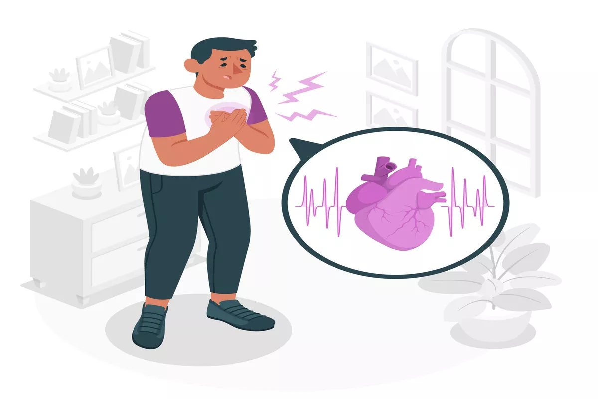

The Cardiology Department is a specialized branch of Aster Whitefield Hospital that focuses on the diagnosis, treatment, and prevention of diseases and disorders related to the cardiovascular system, which includes the heart and blood vessels. It plays an important role in managing various heart conditions and ensuring optimal cardiac health.
The Cardiology Department at Aster Whitefield Hospital has world-class facilities dedicated to providing the highest quality care to patients with cardiovascular disease. Our team of cardiologists, nurses, and technicians has extensive experience and the latest technology to diagnose and treat all types of heart conditions.
We offer a comprehensive range of services including preventive cardiology, interventional cardiology, electrophysiology, echocardiography, and vascular imaging. The Department of Cardiology at Aster Whitefield specializes in the diagnosis and treatment of a wide variety of cardiac conditions, from coronary artery disease to arrhythmias and congestive heart failure. The Department provides a wide array of treatment options for patients with heart conditions. This includes medication management, lifestyle modifications, and various procedures.
Our cardiologists are well-versed in the latest treatment options, including minimally invasive procedures; medication management, lifestyle modification, and more. They are experts and eminent in interventions such as coronary angioplasty, stent placement, cardiac catheterization, pacemaker implantation, cardioversion, and ablation procedures for arrhythmias.
At Aster Whitefield Hospital, we strive to provide our patients with the most advanced care available. We offer a state-of-the-art cardiac catheterization laboratory, with the latest imaging technology and complete interventional services, as well as a complete electrophysiology lab. We strive to provide the best possible care to our patients and believe in an individualized approach to provide personalized attention and optimal results. The Cardiology Department of Aster Whitefield is committed and dedicated to ensuring that all of our patients receive the best possible care in a comfortable and safe environment. We look forward to helping you achieve optimal cardiovascular health.
Our Doctors
We have some of the best specialists from around the world, they bring years of experience and offer evidence-based treatment to ensure the best care for you.


Advanced Technology & Facilities
Well equipped with the latest medical equipment, modern technology & infrastructure, Aster Hospital is one of the best hospitals in India.
Electrocardiography (ECG or EKG)
EKG is an essential diagnostic tool used in cardiology to detect heart conditions and abnormalities. It is a non-invasive and painless procedure that visualizes the heart's electrical activity. Interpretation of an EKG requires specialized training and expertise, and the results can provide valuable information about heart health. It records the heart's electrical activity and produces a visual representation of the heart's rhythm. The EKG machine is composed of electrodes placed on the patient's chest, arms, and legs. These electrodes detect the electromagnetic impulses generated by the heart and transmit them to the EKG machine. This displays the information on a graph. Interpreting an EKG requires specialized training and expertise. A trained healthcare professional, such as a cardiologist or a trained technician, will analyze the EKG results to determine if there are any abnormalities in the heart's rhythm or structure. The results of an EKG can provide valuable information about a patient's heart health and help guide treatment decisions.
Echocardiography-3D
Three-dimensional echocardiography (3D-echo) is a medical imaging technique that allows physicians to visualize the heart in three dimensions. Unlike traditional 2D echocardiography, 3D-echo provides a detailed and comprehensive view of the heart's structure and function. This technique uses specialized ultrasound equipment and software to create a highly detailed 3D image of the heart and its surrounding structures.3D-echo is commonly used to diagnose and monitor a variety of heart conditions, including congenital heart defects, valvular heart disease, and heart failure. By providing a more complete picture of the heart's structure and function, 3D-echo can help physicians make more accurate diagnoses and develop more effective treatment plans.
Transthoracic Echocardiograph
A transthoracic electrocardiogram is a noninvasive method of evaluating the electrical activity of the heart. A chest wall electrode is placed to record the electrical signals produced by the heart. Healthcare professionals can then analyze these signals for abnormalities by amplifying and recording them.
Transthoracic electrocardiograms can provide information regarding a person's heart rhythm, heart rate, and overall health. Abnormalities in the test results may indicate conditions such as arrhythmias, heart attacks, or other cardiac abnormalities. This information can be used to guide further diagnostic testing or to develop a treatment plan for the patient.
3D Transesophageal Echocardiography
Three-D Trans esophageal Electrocardiography is an essential diagnostic tool that helps doctors identify and manage a wide range of heart conditions. It is performed to assess the heart's condition and its functioning. This test is carried out by inserting a probe into the esophagus, allowing a more detailed view of the heart and its structures. During the procedure, the patient is given a sedative to relax. The procedure includes the insertion of a flexible tube with a transducer attached at the end of it. This transducer sends and receives high-frequency sound waves converted into heart images. The procedure usually takes about an hour, and the patient is monitored closely throughout. Three-dimensional Transesophageal Electrocardiography provides detailed information about heart structure and function. It is commonly used to diagnose heart abnormalities, including congenital heart disease, valve disease, and arrhythmias. This test is also used to monitor treatment effectiveness and to evaluate the results of heart surgery.
Dobutamine Stress Echo and Contrast Echocardiography
Dobutamine Stress Echo and Contrast Echocardiography are safe and noninvasive imaging tests used to examine heart conditions. They are performed by trained and skilled healthcare professionals in our Cardiology Team
Dobutamine Stress Echo is a test that combines the use of echocardiography and dobutamine, a medication that increases the heart rate and blood pressure. During the test, the patient is given dobutamine through an IV while an echocardiogram is performed. The test is used to evaluate the function of the heart and to diagnose conditions such as coronary artery disease and heart failure.
Contrast Echocardiography is a type of echocardiogram that uses a contrast agent to enhance the images of the heart. The contrast agent is injected into a vein in the arm and travels to the heart, where it makes the heart chambers and valves easier to see on the echocardiogram. This test is used to evaluate the heart's structure and function and to diagnose conditions such as heart valve disease and congenital heart defects.
Stress testing (exercise or pharmacological)
Stress testing is a valuable tool in the field of cardiology. It is used to evaluate the function of the heart during physical activity. This test is designed to detect any abnormalities or irregularities in the heart's function that may not be evident at rest. The Cardiology Department of Aster Whitefield Hospital provides the facility with exercise stress tests and pharmacologic stress tests to its patients.
Exercise stress tests involve the patient performing physical activity while being monitored to evaluate the heart's response. Pharmacologic stress tests involve the use of medication to increase the heart rate and simulate physical activity.
These stress tests are used by our doctors to diagnose coronary artery disease, a condition where the arteries that supply blood to the heart become narrow or blocked. The test can also be used to assess the severity of heart valve disease and pulmonary hypertension.
Cardiac CT scan
A cardiac CT scan is a non-invasive imaging diagnostic test usually recommended for patients with a high risk of heart disease or those who experience symptoms such as chest pain or shortness of breath. It uses X-ray technology to produce detailed images of the heart and its surrounding blood vessels. The test involves the injection of a contrast dye that highlights the blood vessels and the heart. This dye allows the doctor to see detailed images of the heart and its chambers, as well as any blockages in the coronary arteries. It also allows the doctor to assess the function of the heart valves and detect any signs of heart muscle damage.
Cardiac MRI
Cardiac MRI, also known as magnetic resonance imaging, is a medical imaging technique used to visualize the heart and its surrounding structures. This non-invasive test uses a powerful magnetic field, radio waves, and a computer to generate detailed images of the heart's anatomy and function.
Cardiac MRI is used to diagnose a variety of heart conditions, including heart disease, heart failure, and congenital heart defects. It can also be used to monitor the progression of these conditions over time and to assess the effectiveness of treatment.
Cardiac MRI is a safe and painless procedure that typically takes 30 minutes to an hour. During a cardiac MRI, the patient lies down on a table that slides into a tunnel-like machine. The machine creates a strong magnetic field around the body, which aligns hydrogen atoms. Radio waves are then used to stimulate the hydrogen atoms, causing them to emit signals that are picked up by the machine. These signals are used to create heart images.
The MRI results are reviewed by our expert and qualified radiologists or cardiologist. They can then provide detailed and accurate reports to doctors, helping them to make the best decisions for their patients.
Holter monitoring
It is a valuable tool in diagnosing and treating heart conditions. It provides a detailed analysis of a patient's heart activity over 24 hours, and doctors can make more informed decisions about their treatment plan. This test is typically used to monitor patients with irregular heartbeats or other heart-related symptoms. During the test, a small, portable device called a Holter monitor is attached to the patient's chest with sticky patches.
The Holter monitor continuously records the patient's heart activity for the entire 24-hour period. This allows doctors to analyze the patient's heart rhythms and identify any abnormalities or irregularities.
Patients need to refrain from any conversational behavior during the test. This includes talking directly to others or engaging in activities that may interfere with the accuracy of the test results. Patients should also avoid getting the monitor wet or removing it during the testing period.
Event monitoring (long-term ECG monitoring)
Event monitoring, also known as long-term ECG monitoring, is a diagnostic test that records the electrical activity of the heart over an extended period. This test is used by our cardiologists to diagnose heart rhythm problems that may not be detected during a routine electrocardiogram (ECG) or stress test.
During event monitoring, a small, portable device called a Holter monitor is worn by the patient for up to two weeks. The monitor records the patient's heart rhythm and stores the data for later analysis by a healthcare provider.
Event monitoring is recommended by our doctors often for patients who experience symptoms such as palpitations, dizziness, or fainting spells that may be related to an irregular heart rhythm. The test can help identify the specific type of arrhythmia that is causing the symptoms, allowing for more targeted treatment.
Once the monitoring period is complete, the data collected by the Holter monitor is analyzed by our experts. The results of the test then help us guide treatment decisions and may indicate the need for further testing or intervention.
Ambulatory blood pressure monitoring
Ambulatory blood pressure monitoring (ABPM) is a non-invasive method of monitoring blood pressure over 24 hours. ABPM is useful for people with suspected white coat hypertension, where blood pressure readings are higher in a clinical setting than in everyday life. It can also help diagnose masked hypertension, where blood pressure readings are normal in a clinical setting but higher during daily activities.
It involves wearing a small device connected to a cuff around the upper arm. ABPM is typically done over 24 hours and requires the person to wear the device throughout that time. They are asked to go about their normal daily routine, including sleeping, while the device records their blood pressure. After the monitoring period is complete, the data is downloaded and analyzed by a healthcare provider.
As the device records blood pressure regularly throughout the day and night, it provides a more accurate picture of blood pressure. ABPM results can help our doctors guide treatment decisions, such as adjusting medication dosages or making lifestyle changes to lower blood pressure. It is a vital tool in hypertension management and prevention of associated complications.
TMT treadmill exercise testing
TMT, or treadmill exercise testing, is an important tool used by our cardiologists in the diagnosis and treatment of many heart conditions. These conditions include coronary artery disease, heart failure, and arrhythmias. TMT is used to assess cardiovascular health, detect underlying heart conditions, and evaluate treatment effectiveness.
This non-invasive procedure involves placing the patient on a treadmill and monitoring their heart rate, blood pressure, and ECG while they walk or run at increasing levels of intensity.
During a TMT, the patient is connected to a series of electrodes that measure the heart's electrical activity. The electrodes are attached to the patient's chest, arms, and legs, and are connected to an ECG machine that records the heart's electrical signals. As the patient walks or runs on the treadmill, the intensity of the exercise is gradually increased to assess the heart's response to physical activity.
Tilt table testing
The tilt table test, also known as the "Passive Head-up Tilt Test" or "Head Upright Tilt Test," is a diagnostic procedure used to monitor blood pressure, heart rhythm, and heart rate in a beat-by-beat manner by adjusting the angle of a specialized table on which the patient lies. This test is commonly employed by cardiologists to evaluate patients who frequently experience fainting or exhibit symptoms such as light-headedness.
Fainting medically referred to as syncope, can be caused by various underlying medical conditions involving the heart, nervous system, or inadequate blood flow to the brain.
During a tilt table test, the patient is positioned on a table equipped with a metal footboard to support their feet. Blood pressure cuffs are then placed on one of the patient's arms and a finger, and these cuffs are connected to monitors. The finger cuff continually inflates and deflates, while the arm cuff periodically measures the patient's blood pressure, typically every four to ten minutes.
Electrodes are applied to the patient's chest using adhesive patches and connected to an electrocardiogram (EKG) machine. The EKG records the electrical activity of the heart, which is graphically represented on paper or displayed as a series of lines on a monitor. These lines depict the heart rate and rhythm throughout the test.
All in all, the tilt table test is a valuable tool utilized by our cardiologists to assess patients experiencing episodes of fainting or related symptoms. It involves adjusting the angle of a specialized table while closely monitoring the patient's blood pressure, heart rhythm, and heart rate using cuffs, finger probes, and an electrocardiogram.
FAQs
Want to find out more about the treatment? The answer to your questions can be found below.
What does a Cardiologist do?
A cardiologist examines and evaluates the patient’s cardiovascular system and provides necessary treatment plans according to the patient’s current medical health. They help patients prevent cardiovascular problems, like heart attacks, heart failure, or any kind of heart problem since birth.
What question should you ask a Cardiologist?
Before opting for the cardiologist treatment and surgery, patients must know the side effects of the treatment, precautions required, expected duration of disease curation, specialisation of cardiologists, and their experience. Consult thoroughly to have a better understanding of the best medical treatments.
What are the most common types of conditions that Cardiologists treat?
Expert cardiologists in Whitefield at Aster treat coronary artery disease, heart failure, hypertension, valvular heart disease, congenital heart defects, peripheral artery disease, heart attack, cardiomyopathy, and endocarditis. Consult with cardiologists and experts to know your treatment plan.
When should I consider seeing a Cardiologist?
When you experience symptoms like severe chest pain, dizziness, fainting, palpitations, shortness of breath, swelling in legs, high blood pressure, and diabetic symptoms, they damage blood vessels and heart nerves. Provide complete information to medical staff for effective treatment.
How should I prepare for my first visit to Cardiologist?
Patients must carry their medical history documents, results of recently conducted diagnostic reports, and family heart disease reports and provide complete details if they have high blood pressure, diabetes, or chronic kidney disease. This information helps cardiologists examine patients' health and provide the best treatment plan for them.
How does a Cardiologist decide on a treatment plan?
Cardiologists and medical staff consult with patients regarding available treatment plans and monitor the patient’s and their family's medical history to take a look at any existing genetic conditions that might be affecting their present condition. The cardiologists also assess the possibility of any side effects that may occur as a result of treatment. With the patient’s approval, a treatment plan is put into action.
Will a Cardiologist help manage side effects during treatment?
Yes, trained and specialised cardiologists at Aster thoroughly examine patients’ health progress and diagnostic tests during regular check-ups. This enables them to determine if any side effects have developed as a result of the treatment. They also prevent patients from side effects with the best treatments and care.
What documents should I carry on my first visit to a medical oncologist?
Cardiologists require the patient’s healthcare reports, heart disease reports, diagnostic test results, family heart disease history, and relevant information that influences the treatment plan decisions of expert cardiologists and staff. Detailed patient medical history assists cardiologists in accurately assessing patients' cardiovascular conditions.
How do cardiologists decide if surgery is necessary?
At Aster, expert cardiologists in Bangalore, cardiologists, and medical staff thoroughly study and examine the patient’s medical history and conduct diagnostic tests, stress tests, and cardiac catheterisations. Further, based on the patient’s preferences and medical staff considerations, a treatment decision is formed.
Blogs
The source of trustworthy health and medical information. Through this section, we provide research-based health information, and all that is happening in Aster Hospital.