
New non invasive ultrasound based technology for early detection of liver fibrosis and hepatic steatosis.
Abstract
This article presents two clinical cases of liver diseases commonly seen in day to day practice. First case is 40 yr male asymptomatic patient with cirrhosis of liver. Second case is 30 year female patient with diabetes mellitus and transamnitis. FibroScan of liver is new promising non-invasive, painless, OPD based, affordable method of diagnosing liver fibrosis and liver fat quantification. Early and accurate detection of hepatic fibrosis and hepatic steatosis leads to better treatment outcomes.
Introduction
Liver is one of the vital organs in the body which has unique properties. Firstly development and progress of chronic liver disease (CLD) is asymptomatic in many persons. This leads to delayed diagnosis. Secondly liver has good regenerative capacity which is helpful in liver organ donation in LDLT (Liver donor liver transplantation). Thirdly chronic liver diseases are increasing in recent years due to change in lifestyle, obesity, alcohol abuse. If CLD is detected early in the course of the illness, interventions can be done to halt disease progression.
Case report 1 :
40 year old male patient was referred by his company for annual preventive health check up. He did not give any history of major illness like diabetes mellitus or hypertension, coronary heart disease.
Routine investigations showed:
Hb : 12.3gm%
TLC : 4800/ cmm
Platelet : 1 lakh
LFT : bili (T) : 1.9
SGPT : 70
SGOT : 120
Direct : 1.0 mg%
Indirect : 0.9 mg%
Sr albumin : 3.1
Triple Viral marker : negative
Creatinine : 1 mg%
PT / INR : 18sec / 1.5
USG abdomen showed mild spleenomegaly,no acites ,altered echotexture of liver with normal size . Upper GI scopy showed no esophageal / fundal varices.
On further history taking he gave history of chronic alcohol dependence ( 60 to gms of alcohol per day for last 8 to 10years) . Diagnosis of alcoholic liver disease was made. He was followed for 3 months and advised strict alcohol abstinence.
For further management percutaneous liver biopsy was suggested, but refused liver biopsy as he feels he was asymptomatic. He underwent FibroScan of liver which showed median liver stiffness of 20 KPa with CAP : 270dB/m . Hence diagnosis of CLD : cirrhosis of liver was made. Development of advanced fibrosis correlates with aggressive liver disease in coming years even he was asymptomatic at present.
Case Report 2 :
30 year female patient consulted physician for deranged blood sugars. Her evaluation showed
BMI : 30
Blood sugar fasting : 120
Blood sugar post lunch : 220mg%
HBA1c : 8
Weight : 75 kg
Height : 157 cm
Sr bili(T) : 1.8
SGOT : 70
SGPT : 84
Alkaline phosphate : 98
Sr albumin : 3.8
Triple viral marker : Negative
Creatinine : 0.9 mg%
Sr Triglyceride : 170
Sr LDL : 160 mg%
USG abdomen showed grade I fatty liver. She underwent FibroScan of liver which showed median liver stiffness : 6 KPa with CAP score : 280dB/m suggestive of no fibrosis with S 2 level steatosis .
Discussion on Detection of Liver Fibrosis
Many insults to the liver cause damage to hepatocytes. During process of recovery hepatic stellate cells are activated which cause development of fibrosis. Common insults to the liver are : viruses ( hepatitis B , hepatitis C ) , fatty liver (MAFLD : metabolic associated fatty liver disease) , alcohol , alternative medicines etc . Unless chronic insult stop, hepatic fibrosis continues leading to advanced disease.
Liver biopsy has been gold standard for diagnosing and staging liver fibrosis till few years ago. But due to various factors percutaneous liver biopsy is not done in day to day practice. Since then new technology was required which was easily available, affordable, non invasive for diagnosing liver fibrosis. Radiological and serological methods are available for detecting liver fibrosis. Fibroscan is one of the radiological method. Serum biomarker test measure one or more blood biomarkers and make scoring system. Most commonly used are Hepa score, fibroscore , FIB - 4 score. Studies have shown that these biomarkers test work very well in no/ minimal fibrosis and advanced fibrosis. But in mid level disease their correlation is poor with liver fibrosis. Also radiological and serological markers of fibrosis are not mutually exclusive but complementary, means combing both gives high sensitivity and high specificity.
FibroScan device is research and innovation patented from Echosens ,France. In this technique 50 MHz ultrasound wave probe is used and positioned in right intercostal space near right lobe of liver. Ultrasound wave is passed into the liver and another small transducer at the tip of probe measures the velocity of shear wave (meter/sec) as it passes through the liver. The faster the wave passes through the liver, stiffer the liver. This velocity is converted to liver stiffness as kilopascals by machine software. FibroScan test can be done in almost any patient suspected to have liver disease or used as screening test in health checkup for early detection of CLD.
There are few technical limitations of the test, as it will not give accurate results in patients having ascites , morbid obesity , patients with large chest wall fat . For Obese patient another probe (XL probe) is used.
Discussion on liver fat quantification: (CAP)
With increased prevalence of obesity, there is increase in associated metabolic syndrome with fatty liver disease, diabetes mellitus, and coronary artery disease. Accumulation of fat in the liver was previously thought to be benign and mild disease, but recent evidence suggest it to be significant etiological factor leading to chronic liver disease. Alcohol consumption is associated with simple steatosis in majority majority of people and alcohol abstinence cause disappearance of fat / steatosis.
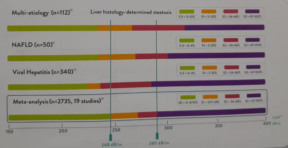
Liver biopsy is gold standard for quanitification of liver fat. Biopsy categorizes fat in liver in three groups namely: S-0 ( no steatosis) , S-1 ( < 33% steatosis) , S-2 ( 34 to 66% steatosis) S-3 ( more than 67% steatosis) . But liver biopsy is not routinely used due to various reasons. Several non invasive methods are used for detecting hepatic steatosis. Commonly used methods are ultrasound, C T scan and MRI of liver. USG is accepted screening method as it is inexpensive with acceptable sensitivity and specificity. But USG interpretation is subjective (changing from person to person) and unable to distinguish steatosis from fibrosis. Non enhanced C T scan has been suggested to quantify > 30 % histological grade hepatic steatosis with 100% specificity and 82% sensitivity. But C T values were significantly different with different manufacture or different generation of C T scan. Besides radiation exposure is a concern.
MRI based technique are sensitive method for detecting steatosis. But are not suitable for routine usage owing to high cost , time consuming method.
Chart showing Cap score correlation with histological liver steatosis .
Controlled attenuation parameter (CAP) used by FibroScan is a novel method for non invasive assessment of steatosis, which measures the attenuation of ultrasound waves when travelling through liver tissue. CAP has multiple advantages: its use is not influenced by liver stiffness, results are immediate, objective (in numbers ) , easy performed method and has good correlation with the amount of steatosis, alcohol use.
Summary :
Fibroscan measurement in the first case clearly shown development of cirrhosis of liver/ advanced fibrosis in asymptomatic patient. FibroScan of liver should be routinely used as screening method in OPD basis set up for early detection of CLD.
Second case showed objective quantification of liver fat by CAP score in FIbroscan test. Successful treatment of hepatic steatosis leads to decrease in CAP score : giving objective results in management of fatty liver disease.