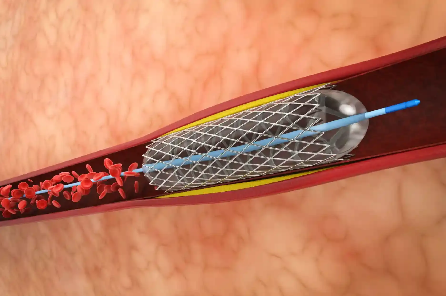

Angioplasty plays a vital role in restoring blood flow by opening blocked coronary arteries. Traditionally, doctors have relied on conventional angiography images to guide stent placement. However, advances in cardiac technology have introduced a more sophisticated approach known as precision angioplasty, which enables cardiologists to treat complex blockages with greater accuracy.
Beyond simply placing a stent, precision angioplasty helps doctors understand the exact nature of a blockage, assess the condition of the artery wall, and manage severe coronary artery disease using advanced imaging and specialised treatment techniques. These innovations are particularly valuable when treating complex blockages and patients requiring urgent cardiac intervention.
What Is Precision Angioplasty?
Conventional coronary angiography provides a two-dimensional (2D) image of the blood vessels, allowing doctors to identify areas of narrowing. However, these images may not always provide sufficient information about the actual dimensions of the artery, the depth of calcium deposits, or the precise characteristics of a blockage.
Precision angioplasty overcomes many of these limitations by incorporating advanced intravascular imaging technologies. These techniques allow cardiologists to examine the internal structure of coronary arteries in much greater detail, helping them select the appropriate stent size, plan treatment more accurately, and assess the results after the procedure.
By enabling a more individualised approach to treatment, precision angioplasty can improve procedural outcomes and reduce certain complications associated with complex coronary interventions.
Looking Inside the Arteries: Intravascular Imaging
Two important imaging technologies used in precision angioplasty are Intravascular Ultrasound (IVUS) and Optical Coherence Tomography (OCT). Both provide detailed information about coronary arteries that may not be visible through conventional angiography alone.
1. Intravascular Ultrasound (IVUS)
IVUS uses high-frequency sound waves to create detailed images of the inside of coronary arteries. A specialised imaging catheter is introduced into the artery, allowing doctors to assess its internal dimensions and the characteristics of the blockage.
IVUS can help cardiologists determine the actual diameter of the artery, evaluate the extent of plaque buildup, identify calcium deposits, and understand the structure of the arterial wall.
This information is particularly useful when treating complex lesions, selecting an appropriately sized stent, and ensuring that the stent expands adequately after placement.
2. Optical Coherence Tomography (OCT)
OCT is an advanced imaging technique that uses near-infrared light to produce highly detailed images of the internal layers of coronary arteries. It offers a higher image resolution than IVUS, allowing doctors to examine small structural details of the arterial wall and plaque.
OCT is especially helpful in assessing the characteristics of a blockage and checking how well a stent has expanded and adapted to the arterial wall after implantation.
By providing detailed information about stent placement and the surrounding tissue, OCT helps cardiologists make informed decisions during complex angioplasty procedures.
Advanced Techniques for Treating Calcified Arterial Blockages
As people age, and particularly among individuals living with long-standing diabetes or kidney disease, calcium deposits can accumulate in the coronary arteries. These deposits can make the arterial walls rigid and difficult to expand.
When severe calcification is present, conventional balloon angioplasty may not adequately open the artery. A stent placed without sufficient preparation of the calcified lesion may fail to expand fully, potentially increasing the risk of future complications, including recurrent narrowing.
To address these challenging situations, cardiologists may use specialised calcium-modification techniques. The choice of procedure depends on the location, severity, and characteristics of the blockage.
1. Rotablation (Rotational Atherectomy)
Rotational atherectomy uses a specialised, diamond-coated burr that rotates at high speed to modify hard, calcified plaque within the coronary artery.
The device selectively breaks down rigid calcium deposits into microscopic particles, helping prepare the artery for balloon expansion and stent placement.
Rotablation can be particularly useful when severe calcification prevents conventional angioplasty equipment from adequately treating the blockage.
2. Intravascular Lithotripsy (IVL)
Intravascular lithotripsy uses a specialised balloon catheter to deliver acoustic pressure waves that modify calcium deposits within the arterial wall.
The technology is based on the principle of lithotripsy, which is also used to break down certain types of kidney stones. In coronary interventions, these pressure waves help fracture deep and superficial calcium deposits, making the artery more flexible and easier to expand.
IVL can help prepare heavily calcified arteries for effective stent placement while limiting damage to surrounding tissues when used appropriately.
3. Orbital Atherectomy (OA)
Orbital atherectomy uses a diamond-coated device that moves in an orbital pattern within the artery to modify calcified plaque.
This technique helps improve the artery's ability to expand and facilitates subsequent stent placement. Its orbital movement allows the device to treat calcified areas of different sizes.
During the procedure, blood flow may continue around the device, although the actual flow depends on the anatomy, device position, and procedural technique.
4. Laser Angioplasty
Laser angioplasty uses specialised laser energy, a process known as photoablation, to break down or remove selected material within a coronary artery.
It may be considered in certain complex situations, including lesions that are difficult to cross with conventional equipment and in-stent restenosis, a condition in which an artery becomes narrowed again within a previously implanted stent.
In selected cases, laser treatment can help modify obstructive tissue and prepare the lesion for further intervention. Its suitability depends on the nature of the blockage and the patient's clinical condition.
Key Benefits of Precision Angioplasty
Precision angioplasty combines detailed imaging with advanced treatment techniques to support more accurate and individualised coronary interventions.
1. More Accurate Stent Placement
Intravascular imaging helps cardiologists measure the artery more precisely and select an appropriate stent size. It also helps assess whether the stent has expanded adequately and is properly positioned against the arterial wall.
Optimal stent expansion is an important factor in achieving good procedural results.
2. Reduced Risk of Certain Complications
By identifying problems such as inadequate stent expansion or incomplete stent apposition, imaging-guided angioplasty allows doctors to correct issues during the procedure.
Appropriate stent sizing and optimisation may help reduce the risk of complications, including stent thrombosis and recurrent narrowing. However, the overall risk also depends on the patient's condition, the complexity of the disease, and adherence to prescribed medication.
3. An Option for Complex Coronary Disease
Patients with multiple coronary artery blockages or extensive calcium deposits may require specialised treatment strategies.
In selected patients, precision angioplasty using intravascular imaging and advanced plaque-modification techniques can provide an alternative to coronary artery bypass surgery. The most appropriate treatment depends on the number and location of the blockages, the complexity of the disease, heart function, associated medical conditions, and the patient's overall health.
A multidisciplinary cardiac team can help determine whether angioplasty, bypass surgery, or another treatment approach is most suitable.
Advances in cardiovascular medicine have made the diagnosis and treatment of coronary artery disease more precise. Precision angioplasty combines advanced imaging and specialised techniques to treat complex blockages and optimise stent placement. While it offers improved treatment options for patients with severe calcification and complex heart disease, careful evaluation by an experienced cardiology team is essential to determine the safest and most appropriate treatment for each patient.