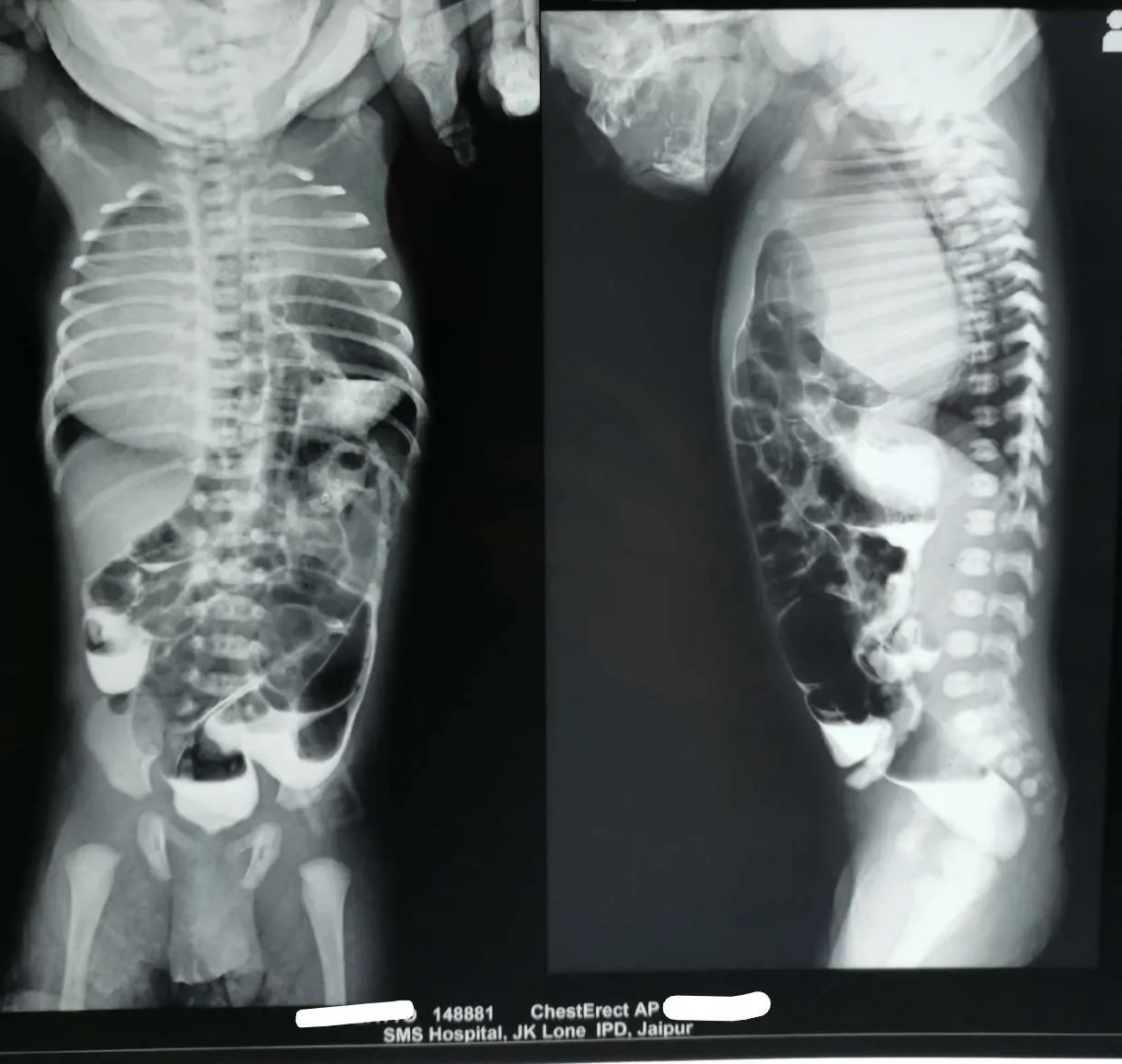

Paediatric surgery as a specialty has undergone a profound paradigm shift over the past few decades. Advances in neonatal intensive care, ultra-fine instrumentation, minimal access techniques, and specialized paediatric anaesthesia have revolutionized patient outcomes. Today, complex congenital conditions that were once associated with significant morbidity in tiny infants are routinely managed with remarkable precision and excellent long-term quality of life.
Despite these landmark advancements, several legacy practices, outdated wait-and-watch mentalities, and clinical misconceptions persist in general practice. Because general practitioners, paediatricians, obstetricians, and emergency physicians form the primary line of contact for young patients, recognizing when to act, when to reassure, and when to refer urgently is paramount.
When evaluating a paediatric surgical problem, the foundational principle remains constant: Children are not miniature adults. Their physiological reserve, fluid dynamics, anatomical proportions, wound healing characteristics, and emotional needs demand age-specific, specialized clinical strategies.
Below is an evidence-based review of some common clinical misconceptions frequently encountered across multi-specialty practice, paired with contemporary surgical realities.
Myth 1: "A non-retractable foreskin in a young boy always requires circumcision or forced manual retraction."
- The Reality: Physiological phimosis is normal in infants and young children; forced retraction causes iatrogenic harm.
- At birth, natural congenital adhesions exist between the inner prepuce and glans, rendering the foreskin non-retractable in over 90% of newborn males. Spontaneous desquamation occurs gradually over time as epithelial debris (smegma) accumulates and intermittent physiological erections separate the tissues. By 3 to 5 years of age, the foreskin becomes naturally retractable in the vast majority of boys without any intervention.
- Clinical Action: Never forcibly retract the foreskin. Forced retraction inflicts painful micro-tears, bleeding, and secondary scarring, ultimately converting a benign, physiological state into true secondary (pathological) phimosis or acute paraphimosis. Reassure anxious parents and reinforce routine exterior hygiene. Circumcision should be reserved for pathological scarring of the preputial tip (Balanitis Xerotica Obliterans - BXO), true ballooning during micturition causing secondary urinary stasis, or recurrent proven UTIs. In non-scarred symptomatic cases, a 2-3 weeks course of topical steroid cream achieves successful resolution in up to 80–90% of boys.
Myth 2: "Wait until the child is 3 to 5 years old before operating on an inguinal hernia or undescended testis."
- The Reality: Delaying surgical correction increases complication rates and permanent tissue damage.
- Inguinal Hernia: Unlike adult direct hernias, paediatric inguinal hernias are virtually always indirect and congenital—resulting from a patent processus vaginalis (PPV). Because the internal ring in infants is narrow and pliable, paediatric hernias carry an exceptionally high risk of incarceration, bowel strangulation, and secondary gonadal ischemia (testicular or ovarian infarction). Repair (herniotomy) should be performed promptly upon diagnosis, regardless of age or body weight.
- Undescended Testis (Cryptorchidism): Progressive, irreversible germ cell degradation and Leydig cell loss begin as early as 6 months of age in an undescended gonad. Contemporary guidelines strongly recommend definitive orchidopexy between 6 to 12 months of age. Deferring surgery beyond the first year of life significantly impairs future fertility potential, increases the risk of testicular torsion, and complicates future surveillance for testicular malignancy.
Myth 3: "Congenital Diaphragmatic Hernia (CDH) is a surgical emergency requiring immediate transfer to the operating room."
- The Reality: CDH is a medical emergency of physiological stabilization, not an immediate surgical emergency.
- Historically, babies with CDH were rushed straight to the operating room for immediate repair. Modern evidence has completely reversed this strategy: mortality in CDH is driven by pulmonary hypoplasia and persistent pulmonary hypertension (PPHN), not the anatomical defect itself.
- Clinical Action: Emergency management focuses on physiological stabilization in the NICU—gentle ventilation strategies (avoiding barotrauma), permissive hypercapnia, inotropic support, and targeted pulmonary vasodilator therapy (e.g., Inhaled Nitric Oxide). Surgical repair of the diaphragm is undertaken only after hemodynamic stability and pulmonary pressures have normalized (typically 24 to 72 hours post-birth). Operating on an unstable infant in persistent pulmonary hypertension carries catastrophic mortality.
Myth 4: "Repeated bilious vomiting in a newborn /infant is simply severe acid reflux, gastritis, or feed intolerance."
- The Reality: Repeated bilious vomiting in a neonate/ infant is a surgical emergency until proven otherwise.
- Bilious fluid indicates an intestinal obstruction distal to the ampulla of Vater. The single most catastrophic entity behind this is Malrotation with Midgut Volvulus. When the gut twists on its narrow mesenteric pedicle, the Superior Mesenteric Artery becomes occluded, leading to total midgut gangrene within hours.
- Clinical Action: Never assume emesis is benign. Do not order trial antacids or feed adjustments. Immediately make the neonate/ infant NPO (nothing by mouth), get a USG abdomen done, place a wide-bore nasogastric tube for active gastric decompression, initiate intravenous fluid resuscitation, and arrange an immediate paediatric surgical consult and upper GI contrast series.
Myth 5: "Intussusception in infants always requires emergency open surgery."
- The Reality: Early diagnosis allows for safe, non-surgical enema reduction; delays lead to gangrene and bowel resection.
- Intussusception (invagination of one segment of bowel into another) is the leading cause of intestinal obstruction in infants aged 3 to 18 months. It classically presents with episodic colicky abdominal pain, drawing up of legs, vomiting, and a palpable sausage-shaped mass (and later, classic "currant jelly" stools).
- The Critical Window: If diagnosed early (typically within 24 to 48 hours of onset without signs of peritonitis or shock), ultrasound-guided hydrostatic (saline) or pneumatic (air) enema reduction succeeds in over 80–90% of cases, completely avoiding surgery. Delaying referral allows progressive venous congestion, ischemia, and bowel necrosis to set in—turning a non-invasive bedside procedure into an emergency laparotomy for gangrenous bowel resection.
Myth 6: "Infantile Hypertrophic Pyloric Stenosis (IHPS) requires immediate operating room transfer."
- The Reality: IHPS is a medical emergency first and a surgical priority second; operating before metabolic resuscitation is unsafe.
- Infants with IHPS (typically 2 to 8 weeks old) present with non-bilious projectile vomiting, dehydration, and a palpable "olive" mass in the epigastrium.
- The Timing Reality: While the diagnosis requires prompt attention, the surgery itself (Ramstedt’s Pyloromyotomy) is never an immediate emergency. Operating on an infant with severe hypochloremic, hypokalemic metabolic alkalosis carries high risks of perioperative cardiac arrhythmias and prolonged post-operative apnea. Dehydration, electrolyte imbalance, and metabolic alkalosis must be fully corrected with IV fluids first over 12–24 hours. Surgery is performed safely once serum electrolytes are within normal limits.
Myth 7: "Neonatal jaundice with pale stools can wait a few months for workup if the baby is active and feeding well."
- The Reality: Biliary Atresia has a strict "Kasai Window"—delaying beyond 60 days dramatically reduces surgical success.
- Persistent conjugated jaundice beyond 2 weeks of life (with acholic/pale stools and dark urine) must be aggressively investigated to rule out Biliary Atresia.
- The Critical Window: The definitive procedure (Kasai portoenterostomy) relies on restoring bile flow before irreversible secondary biliary cirrhosis occurs. If performed before 60 days of life, native liver survival rates exceed 60–70%. If delayed beyond 90 days of life, the operation failure rate is extremely high, leaving liver transplantation as the only option. Never dismiss persistent jaundice as "breast milk jaundice" without checking a fractionated (direct/indirect) serum bilirubin and inspecting stool colour.
Myth 8: "Laparoscopy and minimal access surgery are unsafe or unnecessary for infants and small children."
- The Reality: Paediatric minimal access surgery (MAS) is a well-established gold standard across leading paediatric surgical centres globally.
- With high-definition optics, micro-instrumentation (2mm and 3mm ports), and dedicated paediatric insufflators, advanced laparoscopic and thoracoscopic procedures are routinely performed in neonates weighing under 2.5 kg.
- Procedures such as laparoscopic pyeloplasty, orchidopexy, appendectomy, pull-through operations for Hirschsprung disease, and thoracoscopic repairs of CDH or tracheoesophageal fistula (TEF) offer immense clinical advantages.
- Benefits: Minimal access techniques markedly diminish postoperative pain, reduce surgical site infections, prevent long-term musculoskeletal chest/abdominal wall deformities, decrease hospital stay duration, and provide superior cosmetic results that avoid large growth-restricting abdominal or thoracic scars as the child grows.
Myth 9: "Antenatally detected hydronephrosis usually resolves spontaneously; no formal postnatal evaluation is needed unless symptoms appear."
- The Reality: While many cases are transient, early structured postnatal evaluation is mandatory to safeguard long-term renal nephron mass.
- Antenatal Hydronephrosis (ANH) can represent self-limiting physiological pelvicalyceal ectasia, but it frequently heralds significant pathology—such as severe Pelvi-Ureteric Junction (PUJ) obstruction, Vesicoureteral Reflux (VUR), or life-threatening Posterior Urethral Valves (PUV).
- Reassure parents that minor fluctuations in AP Diameter are normal, avoid repetitive ultrasounds or changing Radiologists, as inter-observer variation creates unnecessary anxiety without altering management.
- Clinical Protocol:
- Bilateral ANH, oligohydramnios, or a solitary kidney: Requires emergency postnatal evaluation (ultrasound within 24–48 hours, bladder catheterization, and early MCU) to exclude lower urinary tract obstruction like PUV before renal decompensation occurs.
- Unilateral ANH: Requires protective prophylactic low-dose antibiotics, a baseline renal ultrasound at 3–7 days of life (avoiding the immediate dehydration phase of the first 48 hours), and specialized nuclear medicine scans (e.g., MAG3 / EC scan) at 4–6 weeks of age.
Myth 10: "An infant or child presenting with acute scrotal pain or swelling should be treated empirically for epididymo-orchitis."
- The Reality: Testicular torsion must be presumed in any acute scrotum until definitively ruled out by USG Doppler study/ surgical exploration.
- True bacterial epididymo-orchitis is exceptionally rare in prepubertal boys unless there is an underlying structural urinary tract malformation or ectopic ureter. Acute scrotal pain, erythema, swelling, or loss of the cremasteric reflex is Testicular Torsion until proven otherwise.
- Golden Window: The warm ischemia time for testicular viability is less than 6 hours. Waiting for an off-hours Doppler ultrasound scan or administering antibiotics frequently delays care past the point of gonadal salvage. If clinical suspicion for torsion is moderate to high, immediate emergency surgical exploration is mandatory—salvaging the testicle takes precedence over diagnostic delay.
Myth 11: "An umbilical hernia requires immediate surgical closure during infancy."
- The Reality: Unlike inguinal hernias, the vast majority of paediatric umbilical hernias undergo spontaneous resolution.
- Paediatric umbilical hernias stem from incomplete fascial closure of the umbilical ring. Unlike inguinal hernias, the fascia around an umbilical defect is smooth and pliable, making acute bowel incarceration exceedingly rare (less than 0.2%). Over 80% to 90% of umbilical hernias close spontaneously by 3 to 4 years of age as the rectus abdominis muscles enlarge and coalesce in the midline.
- Management: Reassure parents and observe. Surgical intervention (umbilical herniorrhaphy) is generally reserved for defects larger than 1.5–2 cm after age 2, symptomatic/painful hernias, incarcerated hernias, or defects that persist beyond 4 years of age
Myth 12: “A congenital hydrocele in an infant requires immediate operation or aspiration."
- The Reality: Paediatric hydroceles are almost always congenital/communicating; most resolve spontaneously, and aspiration is strictly contraindicated.
- Unlike acquired adult hydroceles (which result from fluid imbalance in the tunica vaginalis), a paediatric hydrocele is caused by a persistent, narrow Patent Processus Vaginalis (PPV) allowing peritoneal fluid to track into the scrotum.
- Clinical Action: Spontaneous Resolution: In infants, the PPV frequently obliterates spontaneously within the first year of life as abdominal musculature and tissue rings strengthen. Therefore, simple congenital hydroceles should be observed up to 1 to 2 years of age unless they fluctuate dramatically in size (indicating a wide PPV / frank inguinal hernia).
- Never Aspirate: Needling or aspirating a paediatric hydrocele is dangerous and obsolete. It carries a high risk of bowel perforation (if a hernia sac is present), scrotal infection, and rapid fluid recurrence.
- Surgical Timing: If the hydrocele persists beyond 1.5 to 2 years of age, surgical ligation of the PPV (herniotomy) is indicated.
Conclusion:
Paediatric Surgical care is defined not just by technical skill, but by an in-depth understanding of development physiology, disease specific natural histories and age-critical surgical timing. Managing surgical conditions in children from neonates and infants through to adolescents requires specialised expertise, paediatric surgical management cannot follow one size fits all approach. Every intervention must be carefully individualised based on the child’s age, anatomy and unique physiological reserve. When non specialised management and delayed referral occurs, crucial time windows can slip away, leading to preventable complications. Involving a paediatric surgeon right from the initial presentation ensures that timing is optimized, unnecessary interventions are avoided and children receive the highest standard of dedicated care.