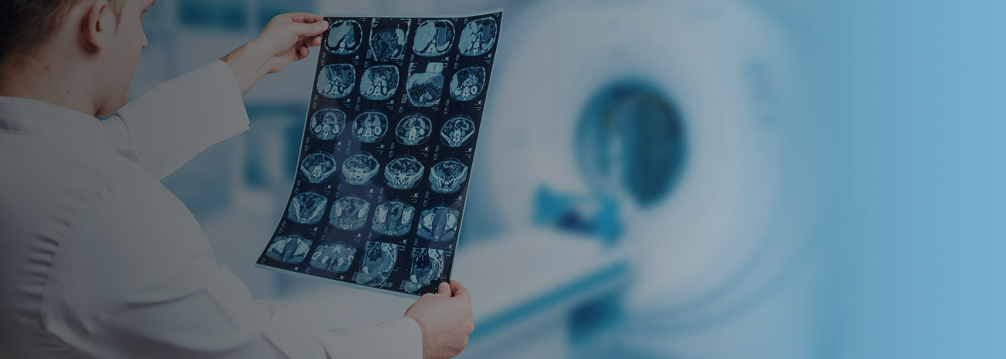
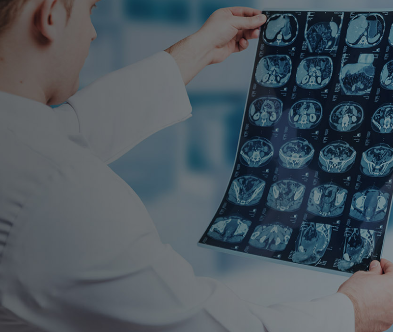
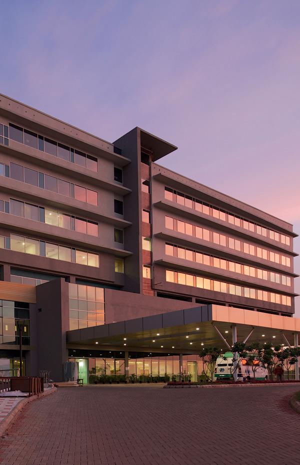
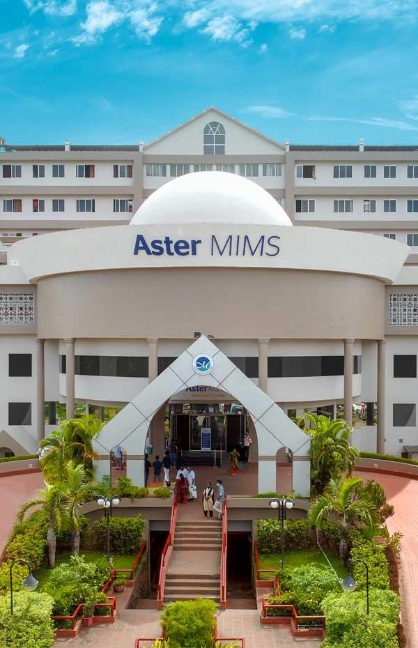
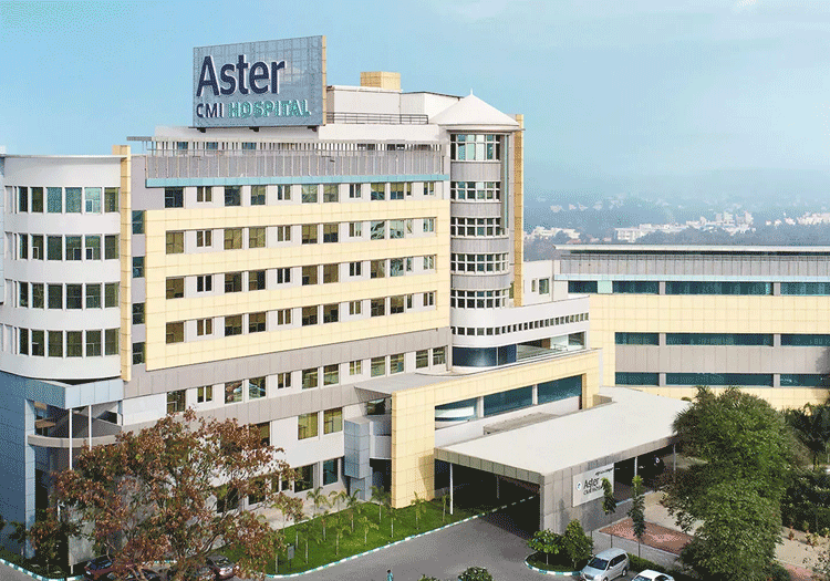
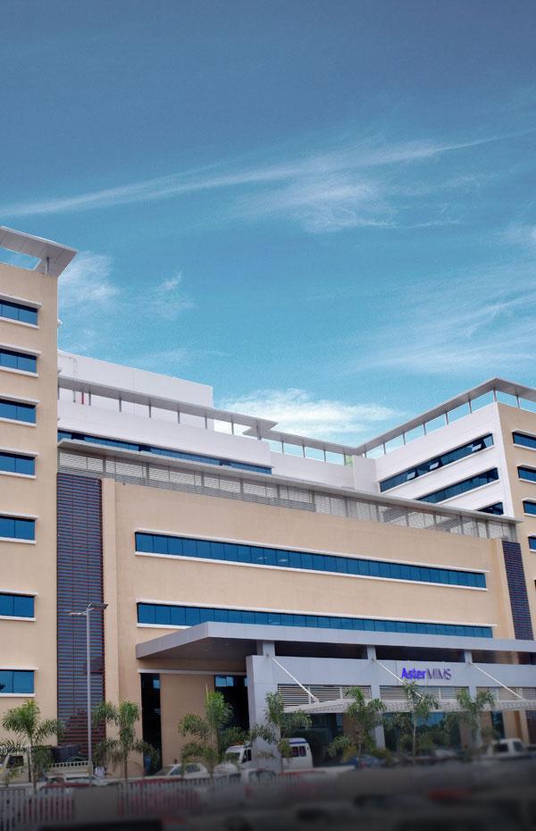
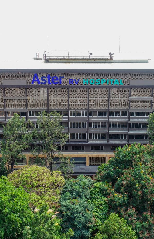
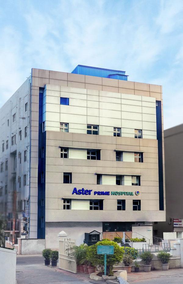
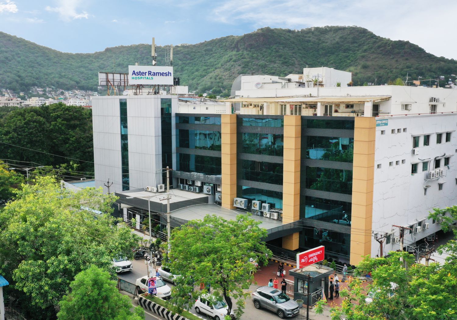
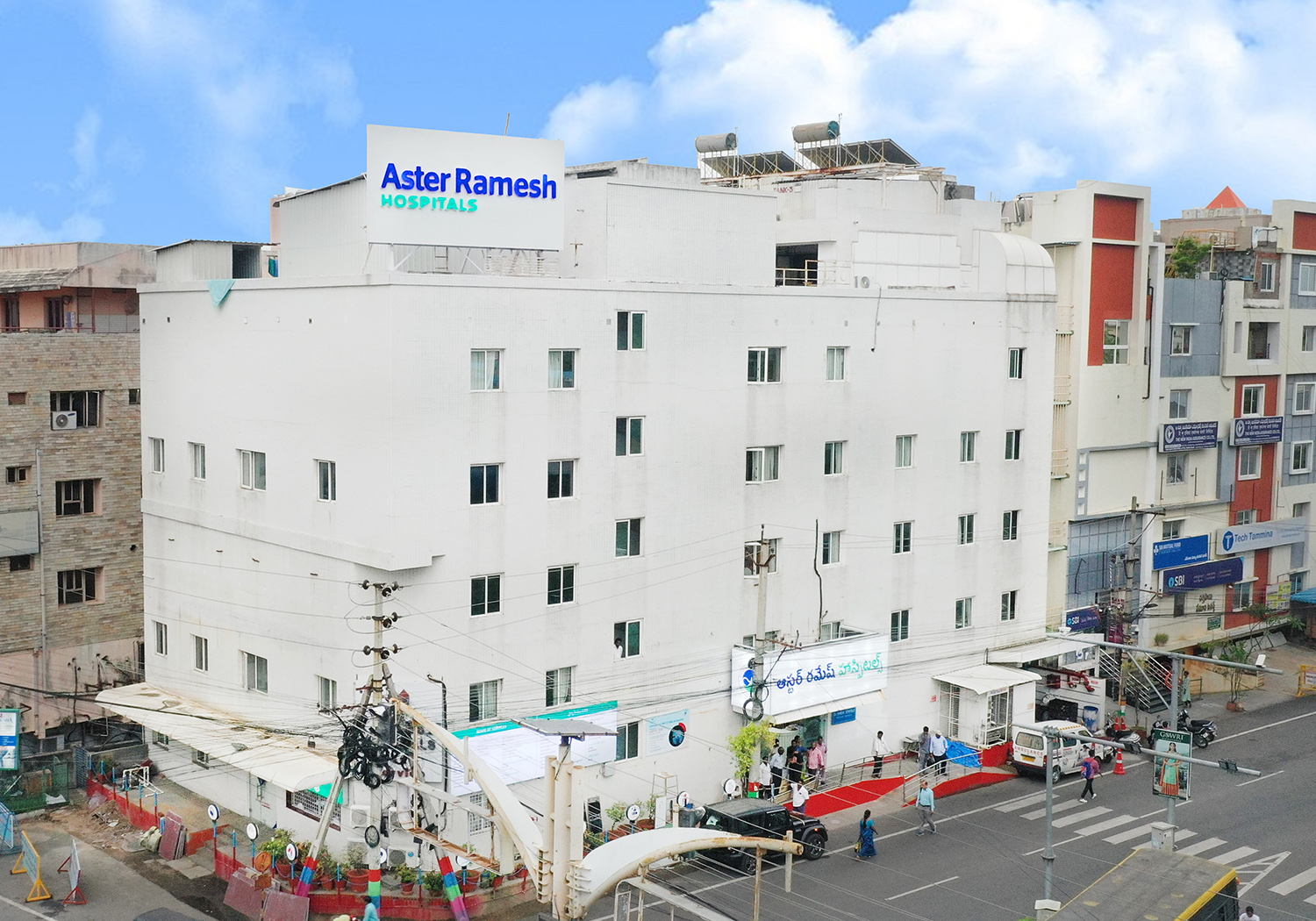
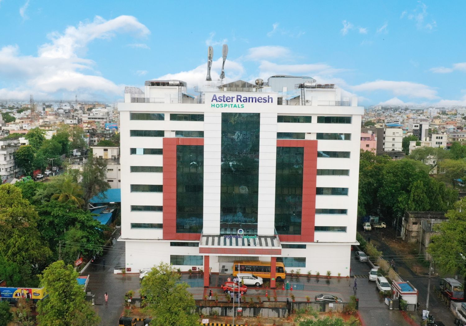
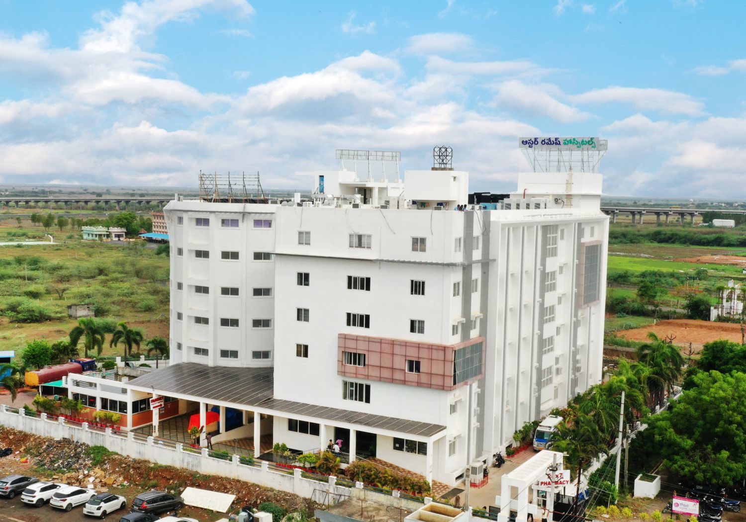
The department of Clinical Imaging & Interventional Radiology uses medical imaging technology to diagnose the disorder stage and prescribe treatment for various medical conditions. At Aster Hospital, we have dedicated radiology unit consisting of a team of experts and well-equipped with modern technologies. The radiology team consists of highly experienced interventional radiologists who have been trained from some of the most prestigious radiology institutes in the world. In addition to diagnostic imaging, our team is also skilled to perform interventional radiological procedures such as administering joint injections, CT and ultrasound-guided radiofrequency ablation, laser ablation, sclerotherapy, tru-cut biopsies, and TRUS guided biopsies. We provide support services for dialysis, endoscopic procedures, OT and critical care procedures, blood bank, neuro-diagnostics and non- invasive cardiology. Our department uses a variety of advanced, high-tech imaging equipment such as MRI scan-1.5 T Philips Achieva, CT scan-16 slice Philips brilliance, Ultrasound-GE Voluson 3D and 4D, Doppler- GE Volluson and BMD-Dexa scan.
Our Doctors
We have some of the best specialists from around the world, they bring years of experience and offer evidence-based treatment to ensure the best care for you.
Treatments & Procedures
We provide comprehensive treatment for all types diseases under one roof. Our highly experienced doctors supported by especially trained clinical staff, ensure the best care for you.
Advanced Technology & Facilities
Well equipped with the latest medical equipment, modern technology & infrastructure, Aster Hospital is one of the best hospitals in India.
Color Doppler is detection and finding motion of blood flow using a Color map that is incorporated standard format B-mode image .It detects and interrogated large scale of region and blood flow, typically blue and red. It can also detected whether the blood moving toward or way from the transducer.It has estimated frequency shift at each point at which motion is detected within an interrogated region, thus yielding information on direction of motion and velocity.
X-rays are a form of electromagnetic radiation, just like visible light. In a health care setting, a machines sends are individual x-ray particles, called photons. These particles pass through the body. A computer or special film is used to record the images that are created.
Multislice CT scanning is a non-invasive medical test that helps physicians diagnose and treat medical conditions. MSCT provide amazing pictures of the human body including 3D images for accurate diagnosis.
In ultrasonography, or ultrasound, high-frequency sound waves, inaudible to the human ear, are transmitted through body tissues.
The echoes are recorded and transformed into video or photographic images.
The Emergency Department has an adjoining Radiology Suite equipped with MRI, Ultrasound, CT Scan and X-ray machine to save on time taken for moving patients to other units/departments for critical diagnostic procedures and fast-tracking the start of treatment.
We have state of art First & Only Philips Brilliance ICT 256 slice CT scanner at Vijayawada Branch and Philips Ingenuity 128 slices low dose CT scanner at Guntur Branch. In addition to routine imaging, we perform brain & neck angiograms, perfusion studies, paediatric and adult coronary angiograms, calcium scoring, EP planning, myocardial perfusion studies, aortograms, pulmonary angiograms, triple rule out studies, CT Bronchoscopy, lung nodule assessment, renal angiograms, liver segmentation planning, upper & lower limb angiograms, dental planning.
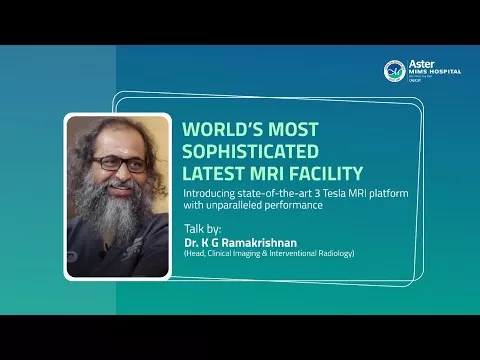
We have state of art First & Only Philips 3 Tesla Ingenia CX Digital MRI, where we can shorten the scan time without compromising image quality. We can perform advanced imaging like neuro perfusion studies, Spectroscopy, subtraction imaging, Cardiac MRI, Foetal MRI, Q flow studies, diffusion weighted whole body imaging with background body signal suppression (DWIBS), Breast MRI, diffusion tensor imaging (DTI), arterial spin labelling (ASL) and Cartilage Mapping.
- US/CT guided FNAC/Biopsy
- Abscess drainages and fluid aspiration with pigtail drainage
- Angiography and venography
- Embolization for PPH, GI bleeders and trauma bleeders
- Embolization in AVM and vascular malformations.
- Thrombolysis – arterial and venous
- Angioplasty and stenting
- Trans-arterial chemotherapy
- Percutaneous biliary procedures including PTBD, Chloecystostomy
- Radiofrequency/ Microwave ablation– liver, lung, renal, bone
- Endovascular aneurysm repair
- Tracheal and esophageal stenting.
- Gastro-intestinal intervention includes TACE, TARE, TIPSS, BRTO, Trans-jugular liver biopsy etc.
- Genito- Urinary interventions include embolization for uterine fibroids, Uterine AVM, benign prostatic hyperplasia, varicocoele, pelvic congestion etc
- Insertion of PICC lines and chemoports.
An advanced Digital Mammography system, it provides high-resolution 3D image results using X-rays for quick detection of even the tiniest calcified lesion that’s in the precancerous stage. The X-ray tubes move in an arc, capturing 11 images in 7 seconds and these images are assembled in the computer to create highly focused 3D image throughout the breast, facilitating easier diagnosis.
The Dexa, utilizing x-ray beams of two different energies, facilitates accurate measurement of bone density and strength with lowest dose of radiation possible, especially for preclinical detection of osteoporosis and preventing complications.
Renders high-resolution radiographs with superior time efficiency. As no repetition and digital enhancement are required, rapid transfer of images for immediate investigation by Physician is made possible through integration with PACS.
Time of Flight PET CT
Accurate and early lesion detection of functional abnormalities and low FDG doses using Time of Flight technology
This Catheterisation Lab (Cath Lab), with the help of Allura Clarity system, reduces radiation doses by 70%, offering significant benefit to the patient as well as staff. Used for Interventional Cardiology and Neurology, the low radiation facilitates repeated investigation if necessary and is also safe for children.
The Biplane Hybrid Cath Lab Unit, which works in a sterile operation theatre environment, combines the traditional diagnostic functions of a Cath lab with surgical functions of an operating room. Designed to undertake complex Neuro vascular and peripheral vascular procedures and Hybrid surgical/ endovascular procedures, the Cathlabs here are the first to have the option of ‘Clarity’, an ingenious system that reduces the radiation dose by upto 70%, which is of significant benefit to the patients. The functions of the unit include:
Biplane Cath Lab
Bi Plane Cath Labs are capable of capturing 3D images in real time, and come with added components like 3D road mapping and post processing to help neurovascular procedures. It can perform interventional procedures in lesser time with lesser usage of contrast, in comparison to single plane.
Digital Subtraction Angiography (DSA)
A type of Fluoroscopy technique used in interventional radiology to clearly visualise blood vessels in a bony or dense soft tissue environment, images are generated using contrast medium by subtracting a ‘pre-contrast image’. Very helpful in diagnosing Neurological problems, the advantage of DSA is that the interference of bony structures can be eliminated.
3D Rotational Angiography
3D rotational angiography provides an image of a particular vessel or chamber while the camera rotates around the patient in a predefined arc, enabling viewing of the structure from multiple angles with a single injection of contrast medium, thereby saving the contrast load and its side effects. A faster procedure compared to conventional Angiography, it is beneficial for patients for whom dye load needs to be restricted and those who require abnormal kidney function test.
CT Like images
With the soft tissue imaging feature that provides enables CT like images, the patient need not to be sent for a CT examination separately. This is a great advantage for neurovascular cases where the mobility of the patient is restricted and timelines are crucial. Interventionist can access CT-like imaging right on the angio system and inturn the soft tissue, bone and other brain structures before, during, or after an interventional procedure. 3D soft tissue imaging supports diagnosis, planning, interventions and treatment follow-up.
Hybrid Cath Lab
In a hybrid application, the Cath lab doubles as an operating room so that a patient undergoing a cardiac cath procedure can have a surgery if required. Enabling the Doctor to address the patient needs quickly, eliminating the need to schedule an additional surgical procedure. As the hybrid cath lab can turn to any angle as required by the interventionist, and can be placed in the corner of the OT when not in use.
Most sophisticated Colour Doppler Systems with Liver Elastography, fusion and navigation for accurate Biopsy, Electronic 4D Imaging with lightweight probes for Cardiac, Radiology and Obstetric Studies. Also provides superior 4D images of the foetus. Department has many state-of-the-art Ultrasound Scanners available for 4D imaging, non-invasive monitoring of liver parenchymal disease with ARFI (Acoustic Radiation Forced Impulse) imaging, with sophisticated image fusion capability to aid accurate guided procedures.
Ultrasound Machines with multi modality image fusion (like CT and MRI) capability and accurate needle tracking.
FAQs
Want to find out more about the treatment? The answer to your questions can be found below.
What Is Interventional Radiology?
A medical subspeciality of Radiology that plays a vital role in both emergency and elective care, Interventional Radiology (IR) is the minimally invasive, image-guided treatment of certain diseases/ conditions that may otherwise require an open surgery.
IR procedures are performed with the help of advanced imaging modalities like MRI, CT and ultrasound scans, in cath labs/ sterile operation theatre environments. The interventional radiologist can see the inside of the body and treat complex conditions ranging from brain aneurysms to cancers, through very small incisions (2-3mm in 90% cases), with unmatched precision and speed.
What are the benefits of Interventional Radiology Procedures
Avoids major surgeries in certain conditions/ diseases
Mostly performed under local anaesthesia/sedation
Tiny incision, minimal scarring
Significantly lesser post-surgery pain
Minimal blood loss
Fewer post-surgical complications
Faster recovery
Shorter hospital stay/ same-day discharge for many procedures
Patient Stories
Our patients are our best advocates, hear the inspiring stories of their treatment journey
Blogs
The source of trustworthy health and medical information. Through this section, we provide research-based health information, and all that is happening in Aster Hospital.
