
Introduction & case summary
Abdomen is temple of surprise and causes of abdominal pain can have wide variety of differentials. Presenting here a case of chronic post prandial abdominal pain that was successfully treated with intervention. A 70 years old gentleman, diabetic & hypertensive for almost three decades presented with abdominal pain for 6 months, pain would start after meals and involve upper and periumbilical region. For this complaints patient visited physicians and specialist. His initial workup involved blood tests and ultrasonography, which were non -diagnostic. Patient received several courses of antacids, probiotics and spasmolytic. Over the period patient underwent upper & lower GI endoscopy which showed gastric ulcers and received symptomatic management. As the symptoms resulted in alteration of food habits and weight loss, a contrast CT of abdomen was eventually done. CECT of abdomen showed diffuse calcification and atherosclerosis of aorta and calcified plaque at origin of common trunk giving origin to celiac artery (CA) and superior mesenteric artery (SMA) – which is an uncommon anatomical variation called celiacomesenteric trunk (CMT). High grade stenosis of this common trunk -celiacomesenteric trunk (CMT) was thought to be the cause of chronic mesenteric ischemia (CMI) resulting in patents symptoms. CA supplies stomach, spleen , liver, duodenum while SMA supplies whole small & part of large intestines- hence very large part of abdominal viscera was ischemic. DSA with intent to perform endovascular management was subsequently planned. DSA showed critical (>90%) ostial stenosis of CMT
Chronic mesenteric ischemia (CMI) is a progressive vascular disorder caused by insufficient blood flow to the bowel, usually due to atherosclerotic narrowing (>90% cases) of the mesenteric arteries. It is often underdiagnosed because symptoms are nonspecific and mimic common gastrointestinal disorders, so a high index of suspicion is needed in patients with postprandial abdominal pain, food fear, and weight loss. In symptomatic patients, multivessel disease is common because extensive collateral circulation often delays symptom onset until at least two major mesenteric vessels are significantly affected. Endovascular therapy, particularly SMA stenting, is now an established treatment option with good symptom relief and lower short-term morbidity than open surgery in many patients.
Incidence and relevance
CMI is rare and accounts for only a small proportion of abdominal pain cases, but it has major clinical consequences when missed. One review noted that atherosclerosis is commonest (>90 %) cause of CMI in adults. The condition is seen more often in elderly patients and has been reported to be more common in women, especially when atherosclerotic risk factors are present. Because the disease often progresses silently until advanced stenosis develops, diagnosis is frequently delayed.
Symptom
Description
Clinical Significance
Postprandial Pain
Abdominal pain occurring 15–30 mins after eating, lasting 1–3 hours
Often epigastric; leads to "food fear" (sitophobia)
Weight Loss
Loss of >5% body weight
Due to voluntary food restriction to avoid pain
Adapted Eating Pattern
Patients eat small, frequent meals or avoid specific foods
A compensatory mechanism to minimize symptoms
Mortality Risk: Untreated symptomatic CMI carries a 5-year mortality rate approaching 100% due to progression to acute mesenteric ischemia (AMI) or severe malnutrition
Causes of ischemia
Mesenteric ischemia can be broadly divided into acute and chronic forms. Chronic disease is most commonly caused by atherosclerotic stenosis or occlusion of the proximal SMA, celiac artery, or both. Less common causes include arterial dissection, vasculitis, fibromuscular dysplasia, radiation injury, arcuate ligament syndrome and cocaine-associated vasospasm or vascular injury. Risk factors mirror systemic atherosclerosis and include smoking, diabetes, hypertension, hyperlipidemia, and generalized peripheral vascular disease.
Diagnostic evaluation
Diagnosis begins with clinical suspicion: postprandial abdominal pain, weight loss, and fear of eating are classic clues. Initial laboratory tests are often nonspecific and may show anemia, electrolyte disturbance, or markers of malnutrition rather than a diagnostic abnormality. CTA is the preferred noninvasive test for confirmation because it defines the degree and location of stenosis and can also assess bowel and collateral circulation. Duplex ultrasound can be used in selected patients, while catheter angiography remains useful when anatomy is unclear or intervention is planned.
Modality
Role
Key Parameters / Findings
CTA (CT Angiography)
Gold Standard for initial diagnosis and pre-procedural planning
Acquisition slice ≤1 mm; arterial + venous/portal phases
CE-MRA
Diagnostic test of choice if CTA contraindicated (e.g., renal failure, contrast allergy)
Contrast-enhanced; avoids iodinated contrast
Duplex Ultrasound
Non-invasive screening; useful for surveillance
SMA PSV ≥275 cm/s or Celiac PSV ≥200 cm/s indicates ≥70% stenosis
Functional Testing
Confirms hemodynamic significance if imaging is equivocal
Measure postprandial flow velocity or lactate levels (less commonly used now)
Endoscopy
Mandatory to exclude GI malignancy
Upper GI endoscopy + colonoscopy (if diarrhea present)
Differential diagnosis
CMI is often mistaken for more common abdominal disorders, so differential diagnosis should be broad. Important alternatives include peptic ulcer disease, chronic pancreatitis, gallstone disease, inflammatory bowel disease, malignancy, functional bowel disorders, and chronic cholecystitis. In older patients with vascular risk factors, recurrent postprandial pain and weight loss should prompt consideration of mesenteric ischemia even when routine GI workup is unrevealing. A parallel evaluation for nutritional compromise and diffuse atherosclerotic disease is also useful.
Management
Once CMI is confirmed, treatment should not be delayed because continued ischemia can progress to acute infarction, perforation, sepsis, and death. Medical therapy alone is generally reserved for patients who are not suitable for revascularization and focuses on risk-factor control and antiplatelet/statin therapy. Revascularization can be open surgical or endovascular, but endovascular stenting is widely favoured in many contemporary cases because of lower early morbidity, shorter hospitalization, and strong symptom relief. After SMA stenting, follow-up with clinical review and serial imaging is important to detect restenosis or recurrence.
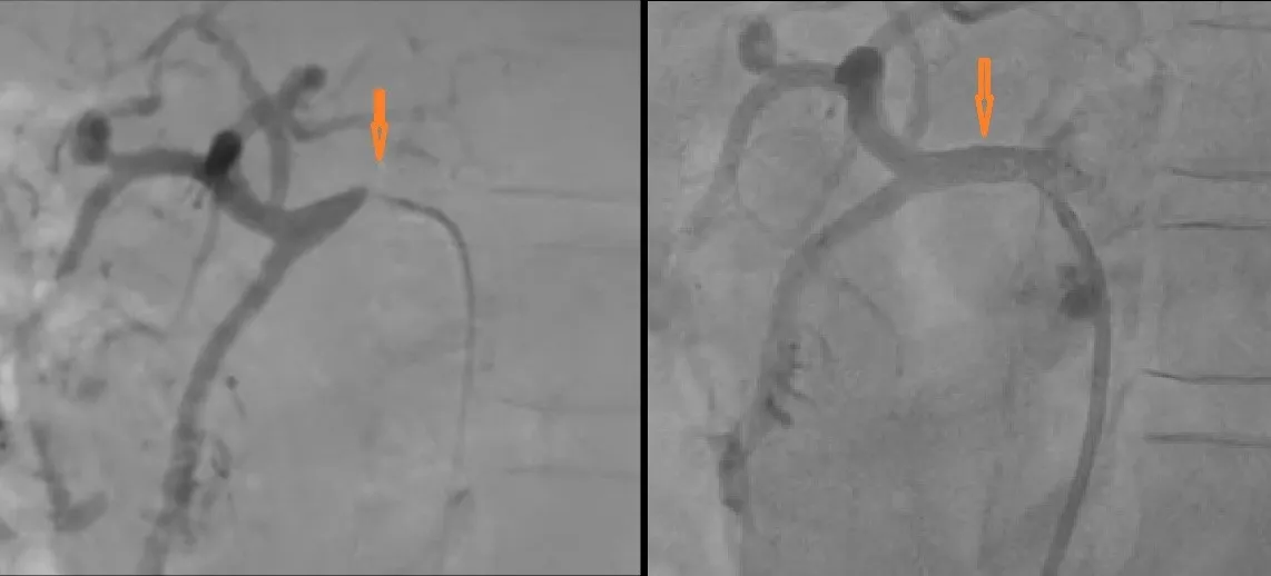
In our present case patient underwent DSA with 5F Cobra C1 catheter. Patient had common origin of celiac and SMA – celiaco-mesenteric trunk which is anatomical variation present in only 0.5-2.5 % of the population -hence large area of gut at ischemic risk. There was significant pressure damping upon engaging CMT with the catheter suggestive of tight calcified ostial stenosis >90% ( Fig 1). Lesion was crossed with guide wire and ostium of CMT was dilated with 4 & 5 x 12 mm NC balloons. A balloon expandable bare metal stent 6 x 15 was deployed at 16 ATM reaching a size of 6.5 mm, ostium of stent was flared with excellent final angiographic result (Fig 2 ). Patient was discharged with significant symptoms relief on dual antiplatelet, statins and other guideline directed treatment.
Discussion
Chronic mesenteric ischemia (CMI) is an uncommon but important cause of postprandial abdominal pain, food fear, and weight loss in older patients with atherosclerotic risk factors. Because symptoms are nonspecific, patients are often first treated for more common gastrointestinal conditions, which delays diagnosis and allows progressive malnutrition and worsening ischemia. This case highlights that a persistent vascular cause should be considered when routine evaluation is unrevealing and symptoms are meal-related.
The unusual and clinically important feature in this patient was the presence of a celiacomesenteric trunk, a rare anatomic variant in which the celiac and superior mesenteric arteries share a common origin. In such anatomy, a single critical ostial lesion can compromise blood flow to both foregut and midgut territories, producing a much larger ischemic burden than isolated stenosis of one vessel. The pressure damping seen during catheter engagement, along with the heavily calcified ostial plaque, strongly supported a hemodynamically significant lesion.
CT angiography is the most useful noninvasive test for suspected chronic mesenteric ischemia because it defines both the vascular lesion and the mesenteric anatomy. In this case, cross-sectional imaging was particularly valuable because it identified the rare common trunk and clarified why the symptom burden was severe despite a relatively nonspecific gastrointestinal workup. Digital subtraction angiography then confirmed the diagnosis and allowed immediate treatment.
Endovascular revascularization is now widely used in chronic mesenteric ischemia because it offers high technical success with lower early morbidity than open surgery. Published series show good symptom relief after stenting, although restenosis and reintervention remain relevant concerns during follow-up. In this patient, balloon predilatation followed by deployment of a balloon-expandable stent at the ostium was appropriate because the lesion was short, calcified, and ostial, where precise positioning and radial force are important.
The choice of a balloon-expandable stent is reasonable in ostial mesenteric disease because it provides accurate placement and strong scaffolding, especially in calcified lesions. The excellent final angiographic result and subsequent symptom relief in this patient support the functional success of the procedure. However, long-term surveillance is still essential because recurrence can occur from in-stent restenosis or progression of diffuse atherosclerotic disease. Global cardiovascular risk reduction is imperative.
This case also reinforces the need for multidisciplinary thinking in chronic abdominal pain. A patient with diabetes, hypertension, weight loss, postprandial pain, and nondiagnostic endoscopy should prompt consideration of mesenteric ischemia even when peptic ulcer disease or other gastrointestinal diagnoses are present. Early recognition is crucial because untreated symptomatic chronic mesenteric ischemia can progress to acute bowel infarction, severe malnutrition, and death.