
Key words: Severe aortic stenosis (AS), Transcatheter aortic valve implantation (TAVI)
Introduction and case summary
Severe calcific aortic stenosis (AS) is an increasingly important cause of heart failure, dyspnea, angina, and reduced functional capacity in octogenarians. In this age group, symptoms are often attributed to other concomitant conditions such as - anemia, renal dysfunction, pulmonary disease, ischemic heart disease or general frailty, which may delay recognition of the valve lesion and obscure its contribution to the patient’s overall clinical deterioration. Once symptoms develop, untreated severe AS carries a poor prognosis, and timely valve should be considered when meaningful survival and quality-of-life improvement are expected.
Transcatheter aortic valve implantation (TAVI) has become an established treatment option for elderly and high-surgical-risk patients, particularly those aged over 80 years when transfemoral access is anatomically feasible. However, decision-making in frail octogenarians with chronic kidney disease, anemia, gastrointestinal bleeding, and concomitant coronary artery disease remains challenging. Such patients require individualized assessment of frailty, procedural risk, renal protection, bleeding risk, anatomical suitability, and patient preference through a multidisciplinary heart-team approach.
Case
An 84-year-old man with bronchial asthma, chronic anemia, chronic kidney disease (baseline serum creatinine 2.0 mg/dL), benign prostatic hyperplasia, and chronic duodenal ulcer was admitted two years earlier with heart failure. Echocardiography demonstrated severe aortic stenosis with preserved left ventricular systolic function. In view of his advanced age and frailty, transcatheter aortic valve implantation (TAVI) was recommended, but the family initially declined the procedure. He was managed conservatively with diuretics and guideline-directed medical therapy for heart failure.
Despite optimal medical treatment, his quality of life remained poor, with recurrent hospitalizations. Six months before the present admission, he developed severe anemia, angina, and dyspnea in the setting of gastrointestinal bleeding. Upper gastrointestinal endoscopy revealed a duodenal ulcer, which was treated with blood transfusions, haematinics, and proton-pump inhibitor therapy. Since he had angina -angiography was performed which showed heavily calcified two-vessel coronary artery disease involving the left anterior descending and right coronary arteries. In view of active gastrointestinal bleeding and the anticipated need for antiplatelet therapy, coronary revascularization was deferred, and the patient was managed medically.
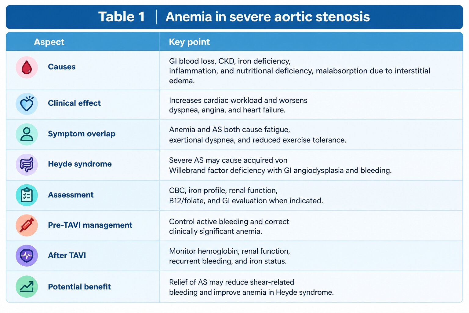
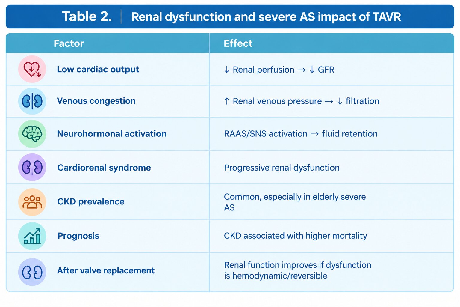
After a few months of clinical stability, he again developed progressive dyspnea and angina on minimal efforts. On readmission, his hemoglobin was 7.0 g/dl and creatinine had risen to 2.4 mg/dl. He received three units of packed red blood cells. Repeat upper gastrointestinal endoscopy showed no active bleeding. Hence the decline was attributable to severe AS itself (Table 1, 2). Echocardiography revealed progression of aortic stenosis, with aortic valve gradients (peak/mean) : 85/ 50 mmHg, and a calculated aortic valve area of 0.6 cm². His multiorgan symptoms were attributed predominantly to heart failure and driving force was severe AS.
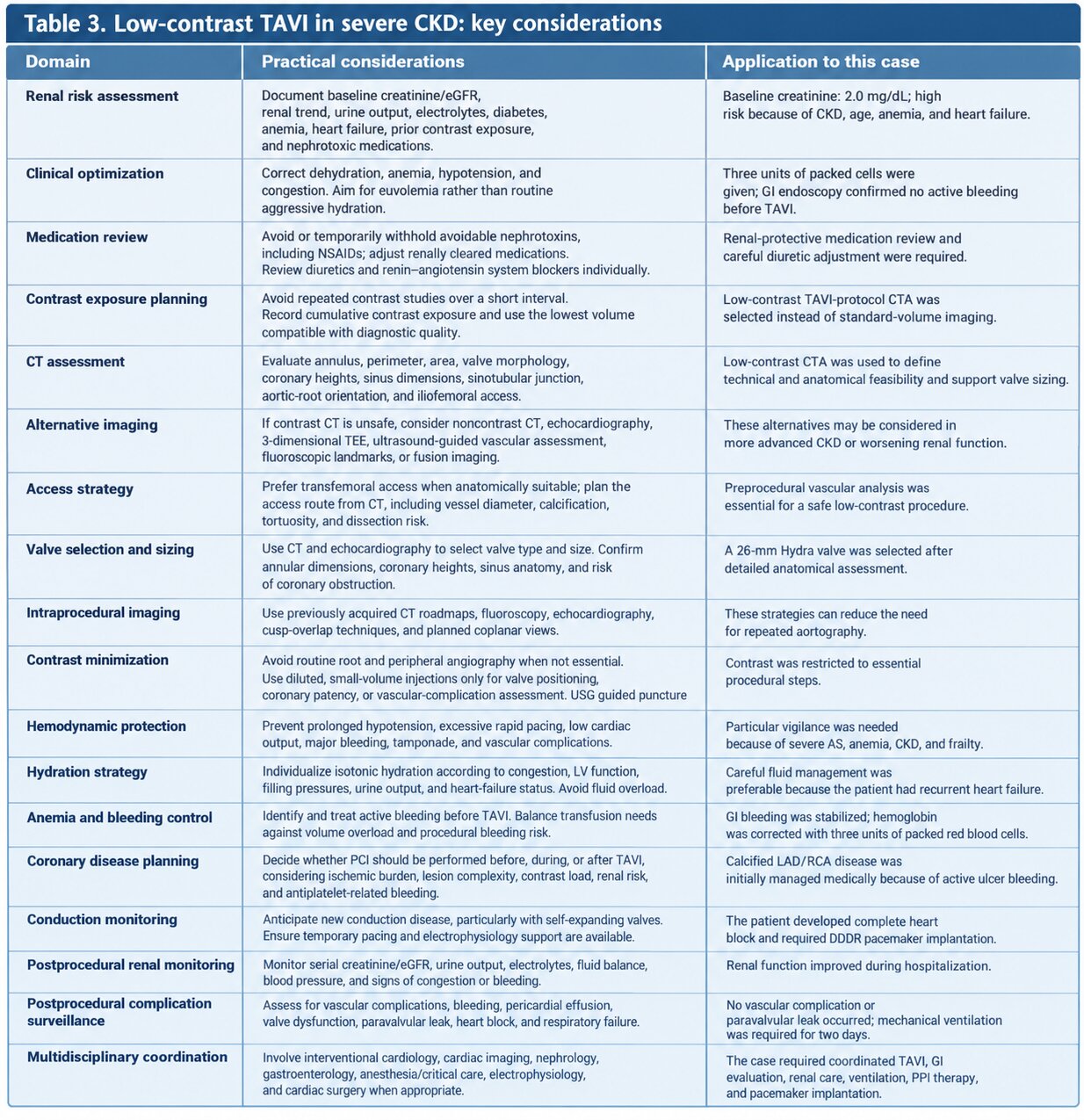
Because of his advanced age, frailty, recurrent heart-failure admissions, chronic kidney disease, severe anemia, and high surgical risk, the heart team reconsidered TAVI. A low-contrast CT angiogram using a dedicated TAVI protocol was performed to assess the aortic annulus, aortic root, coronary artery heights, vascular access, and other anatomical and technical considerations (Table 3). Low-contrast (50 ml- visipaque) planning was selected to minimize the risk of further renal injury, an approach supported by contemporary experience in patients with CKD.
Procedure
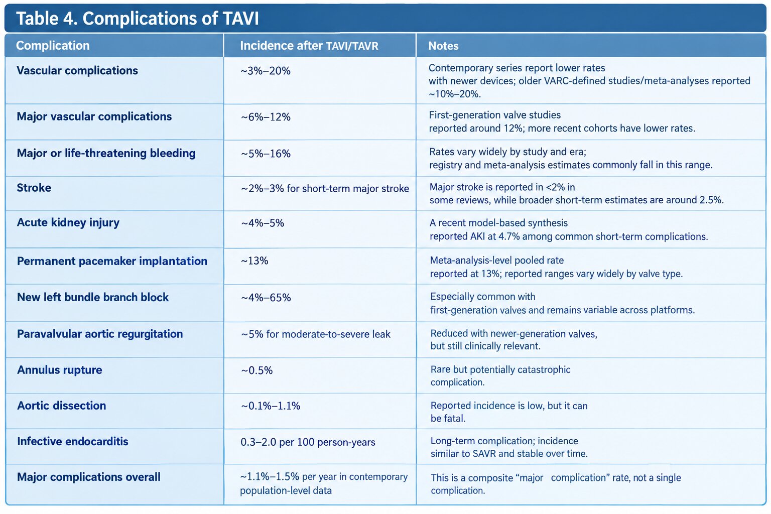
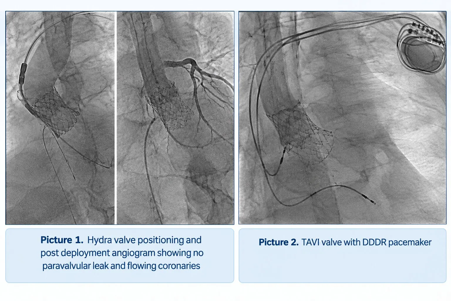
TAVI was performed with 14 French transfemoral access. Valve was predilated with 22 x 40 balloon followed by deployment of a 26-mm Hydra transcatheter heart valve self-expanding valve. There was mild paravalvular leak which was managed with post-dilation with 20 x 40 balloon. Following this, the transvalvular gradients decreased to 12/6 mmHg, with no paravalvular leak or vascular complications. The patient developed complete heart block after the procedure, requiring dual-chamber rate-responsive pacemaker implantation (DDDR) which was performed 2 days later (Picture 1,2). Pacing requirement after TAVI is seen in about 5-10% of patients (Table 4 ).He required mechanical ventilation for two days due to poor respiratory effort and bronchospasm but was subsequently extubated successfully. During the hospital stay, his renal function dramatically improved and creatinine reduced to 1.1 mg/dl & Hb remained stable around 11 g/dl. He was discharged in stable clinical condition.
Discussion
This case demonstrates the central role of severe aortic stenosis in a complex, multimorbid patient. The patient’s recurrent heart-failure admissions, exertional dyspnea, angina, anemia, and renal dysfunction were initially managed as separate clinical problems. However, the clinical course suggested a broader cardiorenal and hemodynamic interaction, with severe aortic stenosis contributing to persistent low functional reserve and repeated decompensation.
Definitive treatment of the valve lesion was associated not only with a marked reduction in transvalvular gradients but also with improvement in renal function and anemia during the hospitalization. These improvements should be interpreted cautiously because the patient also received blood transfusion and treatment for gastrointestinal disease; nevertheless, they support the possibility that relief of severe outflow obstruction improved systemic perfusion and congestion. Renal function after TAVR may remain stable or improve in many patients with CKD, although careful contrast minimization and renal-protective management remain important. Management of concomitant CAD in TAVI patient also needs to be highlighted here. Successful TAVI also improves coronary flow as upstream obstruction gets relived, also contributing to this are reduced LV Filling pressures, reduced sub-endocardial ischemia and improved cardiac output. Thus in most such cases “TAVI first- ischemia guided revascularization later” approach gives adequate symptom relief from CAD point of view
The case also highlights the importance of anticipating conduction complications. Complete heart block is a recognized complication after TAVI and may necessitate permanent pacemaker implantation. In this patient, prompt recognition and dual-chamber pacemaker implantation allowed successful recovery despite the need for temporary mechanical ventilation.
Conclusion
In a frail elderly patient with severe symptomatic aortic stenosis, CKD, chronic anemia, previous gastrointestinal bleeding, and coexisting calcified coronary artery disease, treatment must be individualized and revisited as the clinical condition evolves. A low-contrast TAVI strategy provided definitive treatment of the dominant valvular lesion without vascular complications or paravalvular leak. This case underscores the value of a coordinated heart-team approach involving interventional cardiology, gastroenterology, nephrology, cardiac imaging, anesthesia and critical care, electrophysiology, and nursing teams.
Take-home message
Aortic Stenosis Masquerading as Multimorbidity: The Value of Definitive Intervention
Successful management of complex severe aortic stenosis may require more than valve implantation alone. In carefully selected frail patients, a multidisciplinary, low-contrast TAVI strategy can improve hemodynamics and functional recovery while addressing the interconnected problems of heart failure, renal dysfunction, anemia, gastrointestinal bleeding, and conduction disease.