
Introduction: MI with Non-obstructive Coronaries (MINOCA) is caused by heterogeneous group of coronary artery disorders. MINOCA occurs in 5-15% of patients presenting with acute- ST elevation or non- ST elevation MIs. By angiographic definition, MINOCA have less than 50% diameter stenosis (mild or insignificant coronary stenosis). Commonest coronary causes of MINOCA involve thrombotic lesions caused by plaque ruptures (vulnerable plaques- which are otherwise mild ). Other etiologies involve coronary dissections, spasm, pure thrombus etc. (Fig1). These lesions have significant implications on prognosis and future management. Understanding exact the nature of MINOCA lesions is not always possible on angiography alone. Intra-coronary imaging like OCT gives significant insights on nature and patho-physiology of lesion and aids in decision making. Presenting here two cases of MINOCA where OCT was used for decision making.
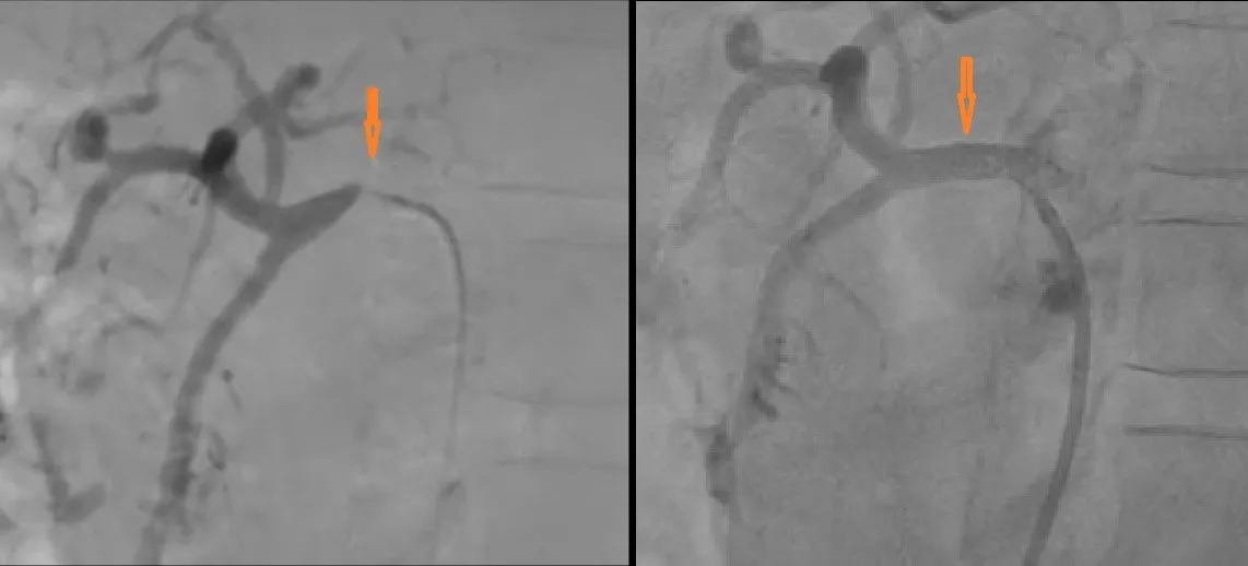
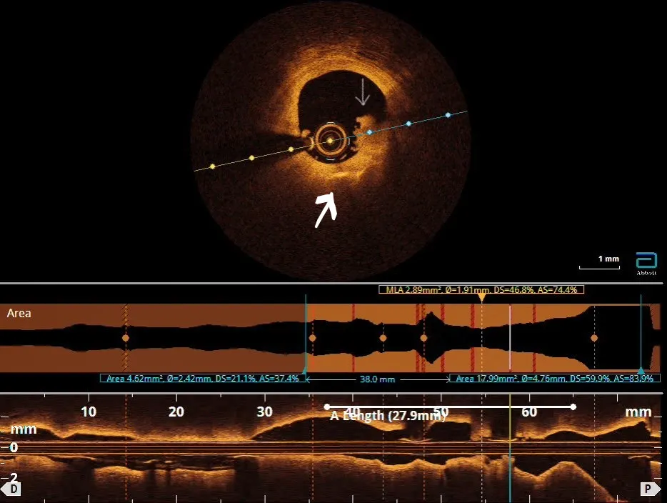
Case 1: 61 years old non-diabetic, hypertensive male had acute Anterior wall MI (AWMI). He was Thrombolysed with Tenectaplase at peripheral center and shifted here for further management. Angiography showed recanalised vessel with Mild Disease in proximal LAD which was less than 50% stenosis (fig2). For mild stenosis, angioplasty (PCI) is not needed but, as he had major AWMI, we decided to do further lesion assessment with OCT study. OCT showed severe fibro-calcific plaque with thrombus (gig3). At tightest lesion part Minimal Luminal Area (MLA) was 2.4 mm2 ( % area stenosis : 83%, % diameter stenosis 60%) ). Total lesion (healthy to healthy landing area) length was 28 mm . As per current OCT guidelines these values were found to be significant and require PCI to improve prognosis and prevent Re-infarction. Adhoc PCI to LAD was done using drug eluting stent 3 x 28 mm size. Post PCI OCT run showed well expanded and apposed stent along its entire length, proximal and distal edge of stent was in healthy area with no edge dissection or thrombus (fig 4). In conclusion, OCT here reclassified the culprit lesion as “significant” which looked mild on angiography. Also Post PCI OCT run confirmed good optimized result post stent.
Case 2 : 33 years old male with past history of cortical venous thrombosis two years back presented with acute AWMI to a peripheral center. He had stopped anticoagulants prescribed by primary care physician. Patient was non diabetic and normotensive. Thrombophilia workup was not done. He was thrombolysed with Reteplase at peripheral center and referred here for angiography and further evaluation. Angiography showed mild disease in proximal LAD with TIMI III flow(fig 5). We decided to do lesion characterization with OCT to guide decision making. OCT showed layered red thrombus in proximal LAD. There was no significant luminal compromise, no significant atherosclerotic component, MLA was …., no dissection or plaque rupture. Based on this data deferred stenting strategy was decided and patient was offered medical management with anticoagulants and antiplatelets drugs. He was later on found to have hyper-homocysteinemia with MTHFR gene mutation heterozygote. Hence long term anticoagulation and folic acid supplementation was given. To conclude, OCT here showed that lesion was in fact a non-occlusive thrombus without any significant atherosclerotic disease hence helping making a decision of deferred stenting with medical anticoagulation for thrombophilic state.
OCT is a versatile intracoronary imaging which helps in lesion characterization, lesion measurements (length, landing zones, %stenosis grading), to decide interventional vs deferred PCI strategy in intermediate lesion, post stenting result optimization, Post PCI complication identification and management.