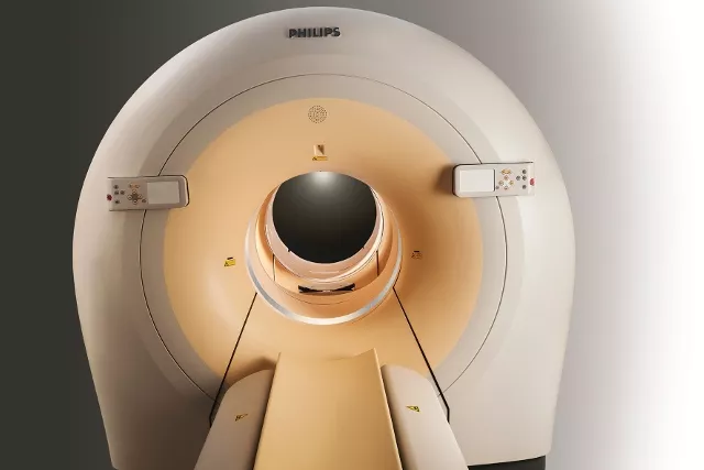
A PET/CT combines two scan techniques in one examination - a PET scan and a traditional CT scan, which is fused using advanced imaging technology and computer software. While some of the other procedures use x-rays or sound waves to create images, a PET scan uses gamma radiation from radioactive tracers to generate 3-dimensional, color images of the functional processes within the human body to detect diseases. By combining these two scanning technologies, a PET/CT scan enables physicians to identify, treat and monitor these diseases earlier and more accurately. This advanced diagnostic service is widely available through experienced Radiologists in Kochi Kerala and leading Radiology Hospital in Kochi Kerala, ensuring accurate and timely diagnosis.
A small amount of radioactive substance or radiotracer similar to glucose, the F 18 fluorodeoxyglucose (FDG) is injected to the patient before the scan. The tracer gives off particles called positrons that release a type of radiation known as gamma rays , which can be detected by the PET scanner. Because cancer cells tend to use more glucose than normal cells, PET scans can help detect the high metabolic activity of these cancer cells to produce images of areas inside the body where glucose is excessively used. These images provide diagnostic information to physicians in treatment planning.
The procedure is painless and takes around 25-30 minutes and does not have any side effects. The radioactive glucose used in nuclear medicine imaging decays quickly and is eliminated through urine.
Preparing for a PET/CT scan:
Before the scan
- Do not eat or drink for at least 4-6 hours before the scan.
- Wear warm comfortable clothing. Avoid wearing jewelry or clothes with metal fasteners or zippers.
- Prescribed medications are fine to be taken on empty stomach.
- Insulin or oral agents used to control diabetes should not be taken within four hours of the PET/CT scan.
- Avoid strenuous exercises on the previous day and on the day of the scan.
After the scan
- Drink plenty of fluids.
- Urinate as often as necessary and empty your bladder frequently for 24 to 48 hours.
- A radiologist will review the images and send a formal report to the patient’s physician within 48 hours.
Today, nuclear medicine imaging offers procedures that are essential in many medical specialties, from pediatrics to cardiology and neurology to oncology. It is used to diagnose and manage the treatment of cancer, heart disease, brain disorders such as Alzheimer’s and Parkinson’s disease, gastrointestinal disorders, lung disorders, bone disorders as well as kidney and thyroid disorders.
It is essential to tell your doctor if you are pregnant or breastfeeding before undergoing a PET scan because of radiation exposure.